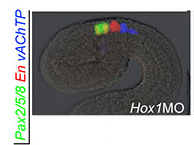
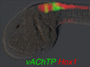
Larvae probed for GFP reporter expression of the promoter region of Ci-vAChTP (named dCi-TPup in the paper).
Original Annotation :
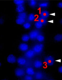
Pictures 1 and 3 : left lateral views, anterior is to the left.
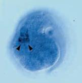
Pictures 2 and 4 : Right lateral views, anterior is to the right.
Picture 5 : Right dorsal view, anterior is to the right.
Pictures 1, 2, 5 : White arrowheads indicate the axon terminals of the cells labeled with asterisks. Some of the labeled cells have an axon with terminal endplates that seem to be neuromuscular junctions (white rectangles). Dotted lines indicate the approximate boundary of the anterior and posterior parts of the visceral ganglion.
Pictures 3, 4 : The white arrowheads indicate three GFP-labeled cells, the white arrows indicate axonal fibers from these cells. The dotted line indicates the trunk-tail boundary.
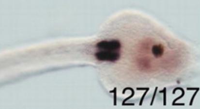
Picture 6 : The green cells in (A) and (B) are GFP-labeled cells, with expression driven by the Ci-vAChTP promoter. The red cells are LacZ-labeled cells with GAD reporter expression.
The red staining in the pics 1, 2, 3, 4 and 5 is due to the UA301 detection. UA301 is a nervous system marker.
In the posterior sensory vesicle, GFP expression driven by the dCi-TPup/GFP transgene was observed. Some extended posteriorly and ended in the middle part of the visceral ganglion.
| Stained molecule |
KH2012:KH.C1.498 (CHAT; CPT2; CRAT) |
| Stained region(s) |
posterior sensory vesicle -
tail nerve cord -
visceral ganglion -
|