Login
Help

GENE CARD

Submit your Data

EST count

27 results

Library Name Percentage Expression
Ciona intestinalis whole animal Ciona intestinalis whole animal 0 clone(s) / 0
Nori Satoh unpublished cDNA library Nori Satoh unpublished cDNA library 1 clone(s) / 5,577
Nori Satoh unpublished cDNA library, young adult Nori Satoh unpublished cDNA library, young adult 0 clone(s) / 31,366
Nori Satoh unpublished cDNA library, cleavage stage embryo Nori Satoh unpublished cDNA library, cleavage stage embryo 0 clone(s) / 15,579
Nori Satoh unpublished cDNA library, egg Nori Satoh unpublished cDNA library, egg 0 clone(s) / 31,100
Nori Satoh unpublished cDNA library, larva Nori Satoh unpublished cDNA library, larva 0 clone(s) / 26,704
Nori Satoh unpublished cDNA library, tailbud embryo Nori Satoh unpublished cDNA library, tailbud embryo 0 clone(s) / 26,680
directional larval cDNA library directional larval cDNA library 0 clone(s) / 39
Ascidian hemocytes cDNA library Ascidian hemocytes cDNA library 0 clone(s) / 0
K. Inaba unpublished cDNA library, testis K. Inaba unpublished cDNA library, testis 0 clone(s) / 5,468
Stratagene UniZAP whole-larva library Stratagene UniZAP whole-larva library 0 clone(s) / 0
Ciona intestinalis larva Ciona intestinalis larva 0 clone(s) / 0
Nori Satoh unpublished cDNA library, blood cells Nori Satoh unpublished cDNA library, blood cells 0 clone(s) / 29,579
Nori Satoh unpublished cDNA library, endostyle Nori Satoh unpublished cDNA library, endostyle 0 clone(s) / 2,556
Nori Satoh unpublished cDNA library, cleaving embryo Nori Satoh unpublished cDNA library, cleaving embryo 0 clone(s) / 16,939
Nori Satoh unpublished cDNA library, gastrula and neurula Nori Satoh unpublished cDNA library, gastrula and neurula 0 clone(s) / 25,258
Nori Satoh unpublished cDNA library, gonad Nori Satoh unpublished cDNA library, gonad 0 clone(s) / 16,936
Nori Satoh unpublished cDNA library, neural complex Nori Satoh unpublished cDNA library, neural complex 0 clone(s) / 10,463
Nori Satoh unpublished cDNA library, heart Nori Satoh unpublished cDNA library, heart 0 clone(s) / 13,243
Yutaka Satou unpublished cDNA library, adult digestive gland Yutaka Satou unpublished cDNA library, adult digestive gland 0 clone(s) / 17,765
Yutaka Satou unpublished cDNA library, embryo whole animal Yutaka Satou unpublished cDNA library, embryo whole animal 0 clone(s) / 17,872
Yutaka Satou unpublished cDNA library, mature adult whole animal Yutaka Satou unpublished cDNA library, mature adult whole animal 1 clone(s) / 107,314
Nori Satoh unpublished cDNA library, juvenile whole animal Nori Satoh unpublished cDNA library, juvenile whole animal 0 clone(s) / 24,372
Nori Satoh unpublished cDNA library, mature adult whole animal Nori Satoh unpublished cDNA library, mature adult whole animal 0 clone(s) / 17,126
Ciona intestinalis whole animal stage3 juvenile Ciona intestinalis whole animal stage3 juvenile 0 clone(s) / 3,798
Yutaka Satou unpublished library (cicx) Yutaka Satou unpublished library (cicx) 0 clone(s) / 2,031
Gateway compatible cien cDNA library, Ciona intestinalis mixed embryonic stages (Egg to Neurula) Gateway compatible cien cDNA library, Ciona intestinalis mixed embryonic stages (Egg to Neurula) 0 clone(s) / 188,431

RNA-Seq transcriptome profiles (by Experiment)

1 result

OVERVIEW

   Expression profile summary from all experiments in WT and mutant conditions:
FPKM & RPKM

Click here to see the RNA-Seq Pipeline Protocol.
You can show/hide lines in the charts by clicking on it in the legend part.
You can see more details by clicking on a point and then, on the link which will appear.

Be careful, FPKM & RPKM does not permit you to compare a gene across different conditions or experiments.

Download FPKM & RPKM data

OVERVIEW

   Expression profile summary from all experiments in WT and mutant conditions:
Relative Log Expression RLE

Be careful, RLE and 2RLE does not permit you to compare a gene across different experiments.

Download RLE Relative Log Expression data

RNA-Seq Experiment n°34135

Read more…

Oocytes of a single individual of Ciona intestinalis (Roscoff, France) were fertilized by a mixture of sperm of two individuals and development was followed microscopically. Embryos were snapfrozen in liquid nitrogen at the appropriate stage and kept at -80°C. Samples at stage 0, 8, 12, 15, 21, 26 were collected from the same fertilzation, while sample from stage 11 was independently collected from a different individual. Total RNA was prepared with the RNA II kit of Magerey Nagel and the quality was checked with the Bioanalyzer, all samples had a RIN value of higher than 9. Strand specific libraries were constructed and the sequencing (50PE) was performed with Illumina HiSeq 2000 technology (BGI, Hong Kong, China). Reads were aligned onto C.robusta genome and considered to reflect the expression of the C.robusta orthologous genes.

Assay platform Illumina HiSeq 2000
Layout Paired-end
Stranded Yes
Read length 49
Normalized data types
  • FPKM
  • RLE
Wild type - Replica 1
Territories whole embryo -
Biomaterial(s) C. robusta formely Ciona int. type A, Stage 0 (Unfertilized egg)
C. robusta formely Ciona int. type A, Stage 8 (64-cell)
C. robusta formely Ciona int. type A, Stage 11 (early gastrula)
C. robusta formely Ciona int. type A, Stage 12 (mid gastrula)
C. robusta formely Ciona int. type A, Stage 15 (mid neurula)
C. robusta formely Ciona int. type A, Stage 21 (mid tailbud I)
C. robusta formely Ciona int. type A, Stage 26 (hatching larva)
Wild type - Replica 2
Territories whole embryo -
Biomaterial(s) C. robusta formely Ciona int. type A, Stage 0 (Unfertilized egg)
C. robusta formely Ciona int. type A, Stage 8 (64-cell)
C. robusta formely Ciona int. type A, Stage 11 (early gastrula)
C. robusta formely Ciona int. type A, Stage 12 (mid gastrula)
C. robusta formely Ciona int. type A, Stage 15 (mid neurula)
C. robusta formely Ciona int. type A, Stage 21 (mid tailbud I)
C. robusta formely Ciona int. type A, Stage 26 (hatching larva)

In Situ Experiment Data

28 results

Experiment data ( results)

C. robusta formely Ciona int. type A, Stage 1 (One cell)

Read more…

Conversion to Aniseed format of data from Yutaka Satou's gene expression profiles of transcription factors and signalling molecule ISH screen (Development. 2004 131:4047-58).

Original annotation :
Gene name :NK6.
Expression pattern : No signal was detected

Stained molecule KH2012:KH.C5.502 (NKX6-1; NKX6-2; NKX6-3)
Stained region(s) No expression -

C. robusta formely Ciona int. type A, Stage 5a (early 16-cell)

Read more…

Conversion to Aniseed format of data from Yutaka Satou's gene expression profiles of transcription factors and signalling molecule ISH screen (Development. 2004 131:4047-58).

Original annotation :
Gene name :NK6.
Expression pattern : No distinct zygotic signal was detected

Stained molecule KH2012:KH.C5.502 (NKX6-1; NKX6-2; NKX6-3)
Stained region(s) whole embryo -

C. robusta formely Ciona int. type A, Stage 6b (late 32-cell)

Read more…

Conversion to Aniseed format of data from Yutaka Satou's gene expression profiles of transcription factors and signalling molecule ISH screen (Development. 2004 131:4047-58).

Original annotation :
Gene name :NK6.
Expression pattern : No distinct zygotic signal was detected

Stained molecule KH2012:KH.C5.502 (NKX6-1; NKX6-2; NKX6-3)
Stained region(s) whole embryo -

C. robusta formely Ciona int. type A, Stage 8 (64-cell)

Read more…

Conversion to Aniseed format of data from Yutaka Satou's gene expression profiles of transcription factors and signalling molecule ISH screen (Development. 2004 131:4047-58).

Original annotation :
Gene name :NK6.
Expression pattern : No distinct zygotic signal was detected

Stained molecule KH2012:KH.C5.502 (NKX6-1; NKX6-2; NKX6-3)
Stained region(s) whole embryo -

C. robusta formely Ciona int. type A, Stage 10 (112-cell)

Read more…

Conversion to Aniseed format of data from Yutaka Satou's gene expression profiles of transcription factors and signalling molecule ISH screen (Development. 2004 131:4047-58).

Original annotation :
Gene name :NK6.
Expression pattern : No distinct zygotic signal was detected

Stained molecule KH2012:KH.C5.502 (NKX6-1; NKX6-2; NKX6-3)
Stained region(s) whole embryo -

C. robusta formely Ciona int. type A, Stage 13 (late gastrula)

Read more…

Conversion to Aniseed format of data from Yutaka Satou's gene expression profiles of transcription factors and signalling molecule ISH screen (Development. 2004 131:4047-58).

Original annotation :
Gene name :NK6.
Expression pattern : No distinct zygotic signal was detected

Stained molecule KH2012:KH.C5.502 (NKX6-1; NKX6-2; NKX6-3)
Stained region(s) whole embryo -

C. robusta formely Ciona int. type A, Stage 15 (mid neurula)

Read more…

Conversion to Aniseed format of data from Yutaka Satou's gene expression profiles of transcription factors and signalling molecule ISH screen (Development. 2004 131:4047-58).

Original annotation :
Gene name :NK6.
Expression pattern : nervous system.

Aniseed annotation :
Later expression is detected in a part of the visceral ganglion, suggesting the two prominent cells are precursors of this later expression territory. The anterior expression in is the central a-line neural plate.

Stained molecule KH2012:KH.C5.502 (NKX6-1; NKX6-2; NKX6-3)
Stained region(s) a line neural plate - row II-B neural plate -

C. robusta formely Ciona int. type A, Stage 17 (initial tailbud I)

Read more…

Conversion to Aniseed format of Ghost database entry from Satou Y et al., Development 2001; 128:2893-2904. Original annotation: . Expression pattern: Epidermis

Stained molecule KH2012:KH.C5.502 (NKX6-1; NKX6-2; NKX6-3)
Stained region(s) epidermis -

C. robusta formely Ciona int. type A, Stage 19 (early tailbud I)

Read more…

Lateral view of the motor ganglion of a WT Ciona robusta early tailbud embryo probed for Nkx6 expression (red) and electroporated with Fgf8/17/18>LacZ reporter construct (green nuclei).
Nkx6 is expressed in A11.117, A11.118 and A11.119 motor neuron precursors (arrowheads).
Original comment: Nuclei of the A9.30 lineage are visualized by immunofluorescence detection of nuclear β-galactosidase reporter (green) driven by the FGF8/17/18 driver (cf diagram).

Stained molecule KH2012:KH.C5.502 (NKX6-1; NKX6-2; NKX6-3)
Stained region(s) visceral ganglion -

C. robusta formely Ciona int. type A, Stage 19 (early tailbud I)

Read more…

Conversion to Aniseed format of Ghost database entry from Satou Y et al., Development 2001; 128:2893-2904. Original annotation: . Expression pattern: Epidermis

Stained molecule KH2012:KH.C5.502 (NKX6-1; NKX6-2; NKX6-3)
Stained region(s) epidermis -

C. robusta formely Ciona int. type A, Stage 19 (early tailbud I)

Read more…

Conversion to Aniseed format of data from Yutaka Satou's gene expression profiles of transcription factors and signalling molecule ISH screen (Development. 2004 131:4047-58).

Original annotation :
Gene name :NK6.
Expression pattern : nervous system, nerve cord.
Comment : cells near the ganglion and a few cells in the brain

Stained molecule KH2012:KH.C5.502 (NKX6-1; NKX6-2; NKX6-3)
Stained region(s) anterior sensory vesicle - visceral ganglion -

C. robusta formely Ciona int. type A, Stage 19 (early tailbud I)

Read more…

Lateral view of an early tailbud wild type embryo probed for Nk6 expression.
Red and yellow arrowheads indicate boundaries between the neck and the visceral ganglion, and between the visceral ganglion and the caudal nerve cord, respectively.

Original Annotation :
Nk6 was expressed in the visceral ganglion (A11.119, A11.118 and A11.117 cell pairs) and in the lateral nerve cord (A11.115 cell pair).

Stained molecule KH2012:KH.C5.502 (NKX6-1; NKX6-2; NKX6-3)
Stained region(s) lateral TNC ependymal precursors - visceral ganglion -

C. robusta formely Ciona int. type A, Stage 19 (early tailbud I)

Read more…

Dorsal view of an early tailbud stage embryo probed for Nk6 expression, following the injection of Pax6 MO.
Red and yellow arrowheads indicate boundaries between the neck and the visceral ganglion, and between the visceral ganglion and the caudal nerve cord, respectively.

Original Annotation :
Expression was lost in the visceral ganglion and in the lateral nerve cord (white arrow).

Stained molecule KH2012:KH.C5.502 (NKX6-1; NKX6-2; NKX6-3)
Stained region(s) No expression -

C. robusta formely Ciona int. type A, Stage 20 (early tailbud II)

Read more…

Lateral view of the motor ganglion of a WT Ciona robusta early tailbud embryo probed for Nkx6 expression (green) and electroporated with FGF8/17/18>mCherry reporter construct (red).
Nkx6 is expressed in A11.119 (arrowhead), A11.118 and A11.119 motor neuron precursors.
The A9.30 lineage is visualized using the FGF8/17/18 reporter construct (red nuclei and diagram).

Stained molecule KH2012:KH.C5.502 (NKX6-1; NKX6-2; NKX6-3)
Stained region(s) visceral ganglion -

C. robusta formely Ciona int. type A, Stage 20 (early tailbud II)

Read more…

Lateral view of the motor ganglion of a Ciona robusta early tailbud embryo probed for Nkx6 expression (green) and electroporated with FGF8/17/18>mCherry reporter construct (red) and FGF8/17/18>Su(H)-DBM overexpression construct.
Nkx6 expression is lost in A11.119 (unfilled arrowhead) but remains in A11.118 and A11.119 motor neuron precursors.
The A9.30 lineage is visualized using the FGF8/17/18 reporter construct (red nuclei).

Stained molecule KH2012:KH.C5.502 (NKX6-1; NKX6-2; NKX6-3)
Stained region(s) visceral ganglion -

C. robusta formely Ciona int. type A, Stage 21 (mid tailbud I)

Read more…

Conversion to Aniseed format of Ghost database entry from Satou Y et al., Development 2001; 128:2893-2904. Original annotation: . Expression pattern: Epidermis

Stained molecule KH2012:KH.C5.502 (NKX6-1; NKX6-2; NKX6-3)
Stained region(s) epidermis -

C. robusta formely Ciona int. type A, Stage 21 (mid tailbud I)

Read more…

Conversion to Aniseed format of data from Yutaka Satou's gene expression profiles of transcription factors and signalling molecule ISH screen (Development. 2004 131:4047-58).

Original annotation :
Gene name :NK6.
Expression pattern : nervous system, nerve cord.
Comment : cells near the ganglion and a few cells in the brain

Stained molecule KH2012:KH.C5.502 (NKX6-1; NKX6-2; NKX6-3)
Stained region(s) anterior sensory vesicle - visceral ganglion -

C. robusta formely Ciona int. type A, Stage 21 (mid tailbud I)

Read more…

Mid tailbud wild type embryo probed for Nk6 expression. No picture available in the article.

Original Annotation :
Nk6 was expressed in the visceral ganglion : A11.118, A12.239, A13.473 cell pairs.

Stained molecule KH2012:KH.C5.502 (NKX6-1; NKX6-2; NKX6-3)
Stained region(s) visceral ganglion -

C. robusta formely Ciona int. type A, Stage 23 (late tailbud I)

Read more…

Conversion to Aniseed format of Ghost database entry from Satou Y et al., Development 2001; 128:2893-2904. Original annotation: . Expression pattern: Epidermis

Stained molecule KH2012:KH.C5.502 (NKX6-1; NKX6-2; NKX6-3)
Stained region(s) epidermis -

C. robusta formely Ciona int. type A, Stage 24 (late tailbud II)

Read more…

Lateral view of the visceral ganglion of a WT Ciona robusta late tailbud embryo electroporated with Nk6>unc76::GFP (green) reporter construct.
Nk6 enhancer drives GFP expression in A11.118 motor neuron precursor.
Original annotation: Nk6>GFP reporter construct labels frondose motor endplates in 50%of electroporated embryos. (A') Magnified view of boxed area in A

Stained molecule KH2012:KH.C5.502 (NKX6-1; NKX6-2; NKX6-3)
Stained region(s) lateral VG ependymal cells -

C. robusta formely Ciona int. type A, Stage 25 (late tailbud III)

Read more…

Dorsal view of the head and neck of a WT Ciona robusta late tailbud embryo electroporated with Vsx>nlsLacZ (red) and probed for Nk6 (KH.5.502) expression (green).
Nk6 is expressed in A11.118 motor neuron precursor and in the epidermis.
Original annotation:) Late Nk6 expression (green) seen in stage E90 embryos in between cells A13.474 and A11.117 as revealed by antibody staining for X-gal in embryos electroporated with Vsx>lacZ.

Stained molecule KH2012:KH.C5.502 (NKX6-1; NKX6-2; NKX6-3)
Stained region(s) head epidermis - lateral VG ependymal cells -

C. robusta formely Ciona int. type A, Stage 26 (hatching larva)

Read more…

Lateral view of the visceral ganglion of a WT Ciona robusta larva co-electroporated with Dmbx>unc76::CFP (blue), Vsx>unc76::mCherry (red), Nk6>unc76::YFP (green), and Islet>unc76::mCherry and Islet>unc76::CFP (purple) reporter constructs.
Dmbx enhancer drives CFP expression in A12.239 motor neuron.
Original annotation:Five motor neurons are simultaneously visualized and distinguishable from one another. Green channel was imaged on a different focal plane than blue or red, to visualize Nk6>YFP-labeled endplates. Yellow color only indicates overlap of cell bodies, not colocalization.
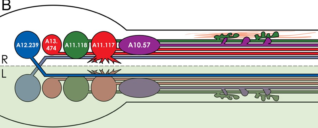
Diagram of the Ciona visceral ganglion ‘brainbow’ showing the neurons from the picture and their unique morphological traits, such as contralateral projection of A12.239 pair, smaller cell body of A13.474, frondose endplates of A11.118, dendrites on A11.117, and elongated cell body and smaller endplates of A10.64 (motor neuron 2). Pale-green shading indicates left side of the larva. Pink fibers represent tail muscle.

Stained molecule KH2012:KH.C1.1212 (DMBX1; DRGX; PITX3)
Stained region(s) motor neurones -

Lateral view of the visceral ganglion of a WT Ciona robusta larva co-electroporated with Dmbx>unc76::CFP (blue), Vsx>unc76::mCherry (red), Nk6>unc76::YFP (green), and Islet>unc76::mCherry and Islet>unc76::CFP (purple) reporter constructs.
Vsx enhancer drives mCherry expression in A13.474 and A11.117 motor neurons.
Original annotation:Five motor neurons are simultaneously visualized and distinguishable from one another. Green channel was imaged on a different focal plane than blue or red, to visualize Nk6>YFP-labeled endplates. Yellow color only indicates overlap of cell bodies, not colocalization.
(B) Diagram showing the neurons from the picture and their unique morphological traits, such as contralateral projection of A12.239 pair, smaller cell body of A13.474, frondose endplates of A11.118, dendrites on A11.117, and elongated cell body and smaller endplates of A10.64 (motor neuron 2). Pale-green shading indicates left side of the larva. Pink fibers represent tail muscle.

Stained molecule KH2012:KH.C11.689 (ALX4; VSX1; VSX2)
Stained region(s) motor neurones -

Lateral view of the visceral ganglion of a WT Ciona robusta larva co-electroporated with Dmbx>unc76::CFP (blue), Vsx>unc76::mCherry (red), Nk6>unc76::YFP (green), and Islet>unc76::mCherry and Islet>unc76::CFP (purple) reporter constructs.
Nk6 enhancer drives YFP expression in A11.118 motor neuron (green).
Original annotation:Five motor neurons are simultaneously visualized and distinguishable from one another. Green channel was imaged on a different focal plane than blue or red, to visualize Nk6>YFP-labeled endplates. Yellow color only indicates overlap of cell bodies, not colocalization.
Diagram of the Ciona visceral ganglion ‘brainbow’ showing the neurons from the picture and their unique morphological traits, such as contralateral projection of A12.239 pair, smaller cell body of A13.474, frondose endplates of A11.118, dendrites on A11.117, and elongated cell body and smaller endplates of A10.64 (motor neuron 2). Pale-green shading indicates left side of the larva. Pink fibers represent tail muscle.

Stained molecule KH2012:KH.C5.502 (NKX6-1; NKX6-2; NKX6-3)
Stained region(s) motor neurones -

Lateral view of the visceral ganglion of a WT Ciona robusta larva co-electroporated with Dmbx>unc76::CFP (blue), Vsx>unc76::mCherry (red), Nk6>unc76::YFP (green), and Islet>unc76::mCherry and Islet>unc76::CFP (purple) reporter constructs.
Islet enhancer drives mCherry and CFP expression in A10.64 motor neuron 2 (purple).
Original annotation:Five motor neurons are simultaneously visualized and distinguishable from one another. Green channel was imaged on a different focal plane than blue or red, to visualize Nk6>YFP-labeled endplates. Yellow color only indicates overlap of cell bodies, not colocalization.
Diagram of the Ciona visceral ganglion ‘brainbow’ showing the neurons from the picture and their unique morphological traits, such as contralateral projection of A12.239 pair, smaller cell body of A13.474, frondose endplates of A11.118, dendrites on A11.117, and elongated cell body and smaller endplates of A10.64. Pale-green shading indicates left side of the larva. Pink fibers represent tail muscle.

Stained molecule KH2012:KH.L152.2 (ISL1; ISL2; LHX9)
Stained region(s) motor neurones -

C. robusta formely Ciona int. type A, Stage 26 (hatching larva)

Read more…

Lateral view of the visceral ganglion of a WT Ciona robusta larva co-electroporated with Nk6>unc76::GFP reporter construct.
Nk6 enhancer drives GFP expression in A11.118 motor neuron.
Original annotation: Some embryos electroporated with NK6>GFP showed frondose endplate labeling by GFP and a single cell body, or soma, strongly labeled by GFP.

Stained molecule KH2012:KH.C5.502 (NKX6-1; NKX6-2; NKX6-3)
Stained region(s) motor neurones -

Lateral view of the visceral ganglion of a WT Ciona robusta larva co-electroporated with Nk6>unc76::GFP reporter construct.
Nk6 enhancer drives GFP expression in A11.118 motor neuron and other cells from the frondose endplate.
Original annotation: Some embryos electroporated with NK6>GFP showed frondose endplate labeling by GFP, but this labeling was associated with multiple labeled cell bodies.

Stained molecule KH2012:KH.C5.502 (NKX6-1; NKX6-2; NKX6-3)
Stained region(s) motor neurones -

C. robusta formely Ciona int. type A, Stage 26 (hatching larva)

Read more…

Dorsal view of the visceral ganglion of a WT Ciona robusta larva co-electroporated with Nk6>unc76::GFP (green) and Dmbx>unc76::mCherry (red) reporter constructs.
Nk6 enhancer drives GFP expression in A11.118 motor neurons.
Original annotation:Co-electroporation of Nk6>GFP and Dmbx>mCherry further supported our conclusion that this late Nk6+ cell is A11.118 and that it is the only motoneuron to form the frondose endplates contacting the lateral surfaces of the anterior tail muscle cells.
(C') Magnified and different focal plane of boxed area to highlight frondose endplate labeled by the Nk6 reporter construct, but not by Dmbx reporter.

Stained molecule KH2012:KH.C5.502 (NKX6-1; NKX6-2; NKX6-3)
Stained region(s) motor neurones -

Dorsal view of the visceral ganglion of a WT Ciona robusta larva co-electroporated with Nk6>unc76::GFP (green) and Dmbx>unc76::mCherry (red) reporter constructs.
Dmbx enhancer drives mCherry expression in A12.239 motor neuron pair.
Original annotation: (C') Magnified and different focal plane of boxed area to highlight frondose endplate labeled by the Nk6 reporter construct, but not by Dmbx reporter.

Stained molecule KH2012:KH.C1.1212 (DMBX1; DRGX; PITX3)
Stained region(s) motor neurones -

C. robusta formely Ciona int. type A, Stage 26 (hatching larva)

Read more…

Dorsal view of the visceral ganglion of a WT Ciona robusta larva co-electroporated with Nk6>unc76::mCherry (green) and Vsx>unc76::GFP (red) reporter constructs. False-color scheme is inverted for consistency in the presentation of the data.
Nk6 enhancer drives mCherry expression in A11.118 motor neurons.
Original annotation: (C') Magnified and different focal plane of boxed area to highlight frondose endplate labeled by the Nk6 reporter construct, but not by Vsx reporter.

Stained molecule KH2012:KH.C5.502 (NKX6-1; NKX6-2; NKX6-3)
Stained region(s) motor neurones -

Lateral view of the visceral ganglion of a WT Ciona robusta larva co-electroporated with Nk6>unc76::mCherry (green) and Vsx>unc76::GFP (red) reporter constructs. False-color scheme is inverted for consistency in the presentation of the data.
Vsx enhancer drives GFP expression in A11.117 and A13.474 motor neurons pairs.
Original annotation: (C') Magnified and different focal plane of boxed area to highlight frondose endplate labeled by the Nk6 reporter construct, but not by Vsx reporter.

Stained molecule KH2012:KH.C11.689 (ALX4; VSX1; VSX2)
Stained region(s) motor neurones -

C. robusta formely Ciona int. type A, Stage 26 (hatching larva)

Read more…

Lateral view of the visceral ganglion of a WT Ciona robusta larva co-electroporated with Nk6>unc76::GFP (green) and Isl>unc76::mCherry (red) reporter constructs.
Nk6 enhancer drives GFP expression in A11.118 motor neurons.
Original annotation:Co-electroporation of Nk6>GFP and Islet>mCherry further supported our conclusion that this late Nk6+ cell is A11.118 and that it is the only motoneuron to form the frondose endplates contacting the lateral surfaces of the anterior tail muscle cells. In fact, co-electroporation with Islet>mCherry suggested that A10.64 might modulate A11.118 presynaptically, by what appears to be contacts onto the frondose endplates themselves.
(C') Magnified boxed area to highlight frondose endplate labeled by the Nk6 reporter construct, but not by Islet reporter.

Stained molecule KH2012:KH.C5.502 (NKX6-1; NKX6-2; NKX6-3)
Stained region(s) motor neurones -

Lateral view of the visceral ganglion of a WT Ciona robusta larva co-electroporated with Nk6>unc76::GFP (green) and Isl>unc76::mCherry (red) reporter constructs.
Islet enhancer drives mCherry expression in A10.64 motor neuron 2.
Original annotation:Co-electroporation of Nk6>GFP and Islet>mCherry further supported our conclusion that this late Nk6+ cell is A11.118 and that it is the only motoneuron to form the frondose endplates contacting the lateral surfaces of the anterior tail muscle cells. In fact, co-electroporation with Islet>mCherry suggested that A10.64 might modulate A11.118 presynaptically, by what appears to be contacts onto the frondose endplates themselves.
(C') Magnified boxed area to highlight frondose endplate labeled by the Nk6 reporter construct, but not by Islet reporter.

Stained region(s) motor neurones -

C. robusta formely Ciona int. type A, Stage 26 (hatching larva)

Read more…

Lateral view of the motor ganglion of a WT Ciona robusta larva electroporated with Nkx6>mCherry reporter construct.
Original comment: Visualization of A11.118-specific frondose endplates (arrowheads) by Nkx6>mCherry. Broad staining in the motor ganglion is due to early Nkx6 throughout the lineage. Nkx6 expression is later restricted to A11.118 motoneuron.

Stained molecule KH2012:KH.C5.502 (NKX6-1; NKX6-2; NKX6-3)
Stained region(s) motor neurones -