Login
Help

GENE CARD

Submit your Data

EST count

27 results

Library Name Percentage Expression
Ciona intestinalis whole animal Ciona intestinalis whole animal 0 clone(s) / 0
Nori Satoh unpublished cDNA library Nori Satoh unpublished cDNA library 1 clone(s) / 5,577
Nori Satoh unpublished cDNA library, young adult Nori Satoh unpublished cDNA library, young adult 0 clone(s) / 31,366
Nori Satoh unpublished cDNA library, cleavage stage embryo Nori Satoh unpublished cDNA library, cleavage stage embryo 0 clone(s) / 15,579
Nori Satoh unpublished cDNA library, egg Nori Satoh unpublished cDNA library, egg 0 clone(s) / 31,100
Nori Satoh unpublished cDNA library, larva Nori Satoh unpublished cDNA library, larva 0 clone(s) / 26,704
Nori Satoh unpublished cDNA library, tailbud embryo Nori Satoh unpublished cDNA library, tailbud embryo 1 clone(s) / 26,680
directional larval cDNA library directional larval cDNA library 0 clone(s) / 39
Ascidian hemocytes cDNA library Ascidian hemocytes cDNA library 0 clone(s) / 0
K. Inaba unpublished cDNA library, testis K. Inaba unpublished cDNA library, testis 0 clone(s) / 5,468
Stratagene UniZAP whole-larva library Stratagene UniZAP whole-larva library 0 clone(s) / 0
Ciona intestinalis larva Ciona intestinalis larva 0 clone(s) / 0
Nori Satoh unpublished cDNA library, blood cells Nori Satoh unpublished cDNA library, blood cells 0 clone(s) / 29,579
Nori Satoh unpublished cDNA library, endostyle Nori Satoh unpublished cDNA library, endostyle 0 clone(s) / 2,556
Nori Satoh unpublished cDNA library, cleaving embryo Nori Satoh unpublished cDNA library, cleaving embryo 0 clone(s) / 16,939
Nori Satoh unpublished cDNA library, gastrula and neurula Nori Satoh unpublished cDNA library, gastrula and neurula 2 clone(s) / 25,258
Nori Satoh unpublished cDNA library, gonad Nori Satoh unpublished cDNA library, gonad 0 clone(s) / 16,936
Nori Satoh unpublished cDNA library, neural complex Nori Satoh unpublished cDNA library, neural complex 0 clone(s) / 10,463
Nori Satoh unpublished cDNA library, heart Nori Satoh unpublished cDNA library, heart 0 clone(s) / 13,243
Yutaka Satou unpublished cDNA library, adult digestive gland Yutaka Satou unpublished cDNA library, adult digestive gland 0 clone(s) / 17,765
Yutaka Satou unpublished cDNA library, embryo whole animal Yutaka Satou unpublished cDNA library, embryo whole animal 1 clone(s) / 17,872
Yutaka Satou unpublished cDNA library, mature adult whole animal Yutaka Satou unpublished cDNA library, mature adult whole animal 0 clone(s) / 107,314
Nori Satoh unpublished cDNA library, juvenile whole animal Nori Satoh unpublished cDNA library, juvenile whole animal 0 clone(s) / 24,372
Nori Satoh unpublished cDNA library, mature adult whole animal Nori Satoh unpublished cDNA library, mature adult whole animal 2 clone(s) / 17,126
Ciona intestinalis whole animal stage3 juvenile Ciona intestinalis whole animal stage3 juvenile 1 clone(s) / 3,798
Yutaka Satou unpublished library (cicx) Yutaka Satou unpublished library (cicx) 0 clone(s) / 2,031
Gateway compatible cien cDNA library, Ciona intestinalis mixed embryonic stages (Egg to Neurula) Gateway compatible cien cDNA library, Ciona intestinalis mixed embryonic stages (Egg to Neurula) 1 clone(s) / 188,431

RNA-Seq transcriptome profiles (by Experiment)

3 results

OVERVIEW

   Expression profile summary from all experiments in WT and mutant conditions:
FPKM & RPKM

Click here to see the RNA-Seq Pipeline Protocol.
You can show/hide lines in the charts by clicking on it in the legend part.
You can see more details by clicking on a point and then, on the link which will appear.

Be careful, FPKM & RPKM does not permit you to compare a gene across different conditions or experiments.

Download FPKM & RPKM data

OVERVIEW

   Expression profile summary from all experiments in WT and mutant conditions:
Relative Log Expression RLE

Be careful, RLE and 2RLE does not permit you to compare a gene across different experiments.

Download RLE Relative Log Expression data

RNA-Seq Experiment n°34135

Read more…

Oocytes of a single individual of Ciona intestinalis (Roscoff, France) were fertilized by a mixture of sperm of two individuals and development was followed microscopically. Embryos were snapfrozen in liquid nitrogen at the appropriate stage and kept at -80°C. Samples at stage 0, 8, 12, 15, 21, 26 were collected from the same fertilzation, while sample from stage 11 was independently collected from a different individual. Total RNA was prepared with the RNA II kit of Magerey Nagel and the quality was checked with the Bioanalyzer, all samples had a RIN value of higher than 9. Strand specific libraries were constructed and the sequencing (50PE) was performed with Illumina HiSeq 2000 technology (BGI, Hong Kong, China). Reads were aligned onto C.robusta genome and considered to reflect the expression of the C.robusta orthologous genes.

Assay platform Illumina HiSeq 2000
Layout Paired-end
Stranded Yes
Read length 49
Normalized data types
  • FPKM
  • RLE
Wild type - Replica 1
Territories whole embryo -
Biomaterial(s) C. robusta formely Ciona int. type A, Stage 0 (Unfertilized egg)
C. robusta formely Ciona int. type A, Stage 8 (64-cell)
C. robusta formely Ciona int. type A, Stage 11 (early gastrula)
C. robusta formely Ciona int. type A, Stage 12 (mid gastrula)
C. robusta formely Ciona int. type A, Stage 15 (mid neurula)
C. robusta formely Ciona int. type A, Stage 21 (mid tailbud I)
C. robusta formely Ciona int. type A, Stage 26 (hatching larva)
Wild type - Replica 2
Territories whole embryo -
Biomaterial(s) C. robusta formely Ciona int. type A, Stage 0 (Unfertilized egg)
C. robusta formely Ciona int. type A, Stage 8 (64-cell)
C. robusta formely Ciona int. type A, Stage 11 (early gastrula)
C. robusta formely Ciona int. type A, Stage 12 (mid gastrula)
C. robusta formely Ciona int. type A, Stage 15 (mid neurula)
C. robusta formely Ciona int. type A, Stage 21 (mid tailbud I)
C. robusta formely Ciona int. type A, Stage 26 (hatching larva)

RNA-Seq Experiment n°34138

Read more…

Ciona robusta adults were obtained from the National Bio-Resource Project for Ciona (NBRP, Japan). 50 unperturbed and Foxd-morphant embryos were collected at the 32-, 64-, and 112-cell stages. RNA was extracted using a Dynabeads mRNA DIRECT Purification Kit (Thermo Fischer Scientific) and libraries were made with an Ion Total RNA-Seq kit ver 2 (Thermo Fischer Scientific). The libraries were sequenced with an Ion PGM instrument (Thermo Fischer Scientific).

Assay platform Ion Torrent PGM
Layout Single-end
Stranded Yes
Read length 20-365
Normalized data types
  • RPKM
Wild type
Territories whole embryo -
Biomaterial(s) C. robusta formely Ciona int. type A, Stage 6a (early 32-cell)
C. robusta formely Ciona int. type A, Stage 8 (64-cell)
C. robusta formely Ciona int. type A, Stage 10 (112-cell)
Mutant
Deregulated molecule(s) KH2012:KH.C8.890 -
Territories whole embryo -
Biomaterial(s) C. robusta formely Ciona int. type A, Stage 6a (early 32-cell)
C. robusta formely Ciona int. type A, Stage 8 (64-cell)
C. robusta formely Ciona int. type A, Stage 10 (112-cell)

RNA-Seq Experiment n°34139

Read more…

The purpose of this project is to identify genes positively and negatively regulated by signaling molecules that belongs to the TGFbeta superfamily.

Assay platform Illumina HiSeq 2500
Layout Single-end
Stranded Yes
Read length 101
Normalized data types
  • RPKM
Wild type
Territories whole embryo -
Biomaterial(s) C. robusta formely Ciona int. type A, Stage 13 (late gastrula)
Mutant 1
Deregulated molecule(s) KH2012:KH.C4.125 -
Territories whole embryo -
Biomaterial(s) C. robusta formely Ciona int. type A, Stage 13 (late gastrula)
Mutant 2
Deregulated molecule(s) KH2012:KH.C2.573 -
Territories whole embryo -
Biomaterial(s) C. robusta formely Ciona int. type A, Stage 13 (late gastrula)

In Situ Experiment Data

96 results

Experiment data ( results)

C. robusta formely Ciona int. type A, Stage 1 (One cell)

Read more…

Conversion to Aniseed format of data from Yutaka Satou's gene expression profiles of transcription factors and signalling molecule ISH screen (Development. 2004 131:4047-58).

Original annotation :
Gene name :Neurogenin.
Expression pattern : No signal was detected

Aniseed annotation :
A very weak staining is observed through the embryo (maternal signal?)

Stained molecule KH2012:KH.C6.129 (NEUROG1; NEUROG2; NEUROG3)
Stained region(s) whole embryo -

C. robusta formely Ciona int. type A, Stage 5a (early 16-cell)

Read more…

Conversion to Aniseed format of data from Yutaka Satou's gene expression profiles of transcription factors and signalling molecule ISH screen (Development. 2004 131:4047-58).

Original annotation :
Gene name :Neurogenin.
Expression pattern : No distinct zygotic signal was detected

Aniseed annotation :
A very weak staining is observed through the embryo (maternal signal?)

Stained molecule KH2012:KH.C6.129 (NEUROG1; NEUROG2; NEUROG3)
Stained region(s) whole embryo -

C. robusta formely Ciona int. type A, Stage 6b (late 32-cell)

Read more…

Conversion to Aniseed format of data from Yutaka Satou's gene expression profiles of transcription factors and signalling molecule ISH screen (Development. 2004 131:4047-58).

Original annotation :
Gene name :Neurogenin.
Expression pattern : A6.1, A6.3, B6.1

Stained molecule KH2012:KH.C6.129 (NEUROG1; NEUROG2; NEUROG3)
Stained region(s) A6.1 cell pair - A6.3 cell pair - B6.1 cell pair -

C. robusta formely Ciona int. type A, Stage 8 (64-cell)

Read more…

Conversion to Aniseed format of data from Yutaka Satou's gene expression profiles of transcription factors and signalling molecule ISH screen (Development. 2004 131:4047-58).

Original annotation :
Gene name :Neurogenin.
Expression pattern : No distinct zygotic signal was detected

Aniseed annotation :
A very weak staining is observed through the embryo

Stained molecule KH2012:KH.C6.129 (NEUROG1; NEUROG2; NEUROG3)
Stained region(s) whole embryo -

C. robusta formely Ciona int. type A, Stage 10 (112-cell)

Read more…

Conversion to Aniseed format of data from Yutaka Satou's gene expression profiles of transcription factors and signalling molecule ISH screen (Development. 2004 131:4047-58).

Original annotation :
Gene name :Neurogenin.
Expression pattern : A8.16.

Stained molecule KH2012:KH.C6.129 (NEUROG1; NEUROG2; NEUROG3)
Stained region(s) A8.16 cell pair -

C. robusta formely Ciona int. type A, Stage 10 (112-cell)

Read more…

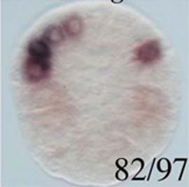
Vegetal pole view of a 110-cell stage wild type embryo probed for Ci-neurogenin expression. Ci-neurogenin expression was restricted to the lateral-most-A-line neural blastomere, A8.16 cell pair. Numbers indicate any expression other the total number of embryos analysed.

Stained molecule KH2012:KH.C6.129 (NEUROG1; NEUROG2; NEUROG3)
Stained region(s) A8.16 cell pair -

C. robusta formely Ciona int. type A, Stage 10 (112-cell)

Read more…

Vegetal pole view of a 110-cell stage embryo probed for Ci-Ngn (neurogenin) expression in which Nodal signaling has been blocked with SB431542. Inhibition of the Nodal signaling blocked Ci-Ngn expression in the A8.16 cells. Numbers indicate any expression over the total number of embryos analysed.

Stained molecule KH2012:KH.C6.129 (NEUROG1; NEUROG2; NEUROG3)
Stained region(s) No expression -

C. robusta formely Ciona int. type A, Stage 10 (112-cell)

Read more…

Vegetal pole view of a 110-cell stage embryo probed for Ci-Ngn (neurogenin) expression in which Nodal signaling has been blocked with Nodal-Mo. Inhibition of the Nodal signaling blocked Ci-Ngn expression in the A8.16 cells. Numbers indicate any expression over the total number of embryos analysed.

Stained molecule KH2012:KH.C6.129 (NEUROG1; NEUROG2; NEUROG3)
Stained region(s) No expression -

C. robusta formely Ciona int. type A, Stage 10 (112-cell)

Read more…

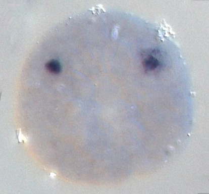
Vegetal view of a 110-cell stage wild type embryo probed for Ci-neurogenin expression. Ci-neurogenin expression was detected in A8.16 cell pair. Numbers indicate any expression over the total number of embryos analysed.

Stained molecule KH2012:KH.C6.129 (NEUROG1; NEUROG2; NEUROG3)
Stained region(s) A8.16 cell pair -

C. robusta formely Ciona int. type A, Stage 10 (112-cell)

Read more…

Vegetal view of an early gastrula stage probed for Ci-neurogenin expression following the injection of Delta2-MO. Numbers indicate any expression over the total number of embryos analyzed

Original Annotation : Ci-neurogenin expression was blocked by injection of Delta2-MO (inhibition of Delta2/Notch signaling).

Stained molecule KH2012:KH.C6.129 (NEUROG1; NEUROG2; NEUROG3)
Stained region(s) No expression -

C. robusta formely Ciona int. type A, Stage 10 (112-cell)

Read more…

Vegetal view of an early gastrula stage probed for Ci-neurogenin expression following DAPT injection. Numbers indicate any expression over the total number of embryos analyzed

Original Annotation : Ci-neurogenin expression was blocked by the inhibition of Delta2/Notch signaling due to DAPT.

Stained molecule KH2012:KH.C6.129 (NEUROG1; NEUROG2; NEUROG3)
Stained region(s) No expression -

C. robusta formely Ciona int. type A, Stage 10 (112-cell)

Read more…

Vegetal view of an early gastrula stage probed for Ci-neurogenin expression following the injection of pFOG::Delta2 construct. Numbers indicate any expression over the total number of embryos analyzed

Original annotation : overexpression of Delta2 produced ectopically expression of Ci-neurogenin in A8.15, A8.8, A8.7 cell pairs.

Stained molecule KH2012:KH.C6.129 (NEUROG1; NEUROG2; NEUROG3)
Stained region(s) A8.15 cell pair - A8.16 cell pair - A8.7 cell pair - A8.8 cell pair -

C. robusta formely Ciona int. type A, Stage 10 (112-cell)

Read more…

Graph showing the percentage (0 to 100, on the y axis) of early gastrula embryos with strong or weak Ci-neurogenin expression in the lateral neural precursors (arrow in the picture) following SB431542 treatment at the 16-cell stage."C" means control embryos (133 embryos probed). 'Strong' means at least one cell stained like in the control picture, and 'weak' means at least one blastomere showing very weak expression.

Original Annotation : there is no embryo with Ci-neurogenin expression when the embryos were treated with SB431542 at 16-cell stage. In the control A8.16 cell pair is stained.

Stained molecule KH2012:KH.C6.129 (NEUROG1; NEUROG2; NEUROG3)
Stained region(s) No expression -

C. robusta formely Ciona int. type A, Stage 10 (112-cell)

Read more…

Graph showing the percentage (0 to 100, on the y axis) of early gastrula embryos with strong or weak Ci-neurogenin expression in the lateral neural precursors (arrow in the picture) following SB431542 treatment at the 32-cell stage."C" means control embryos (133 embryos probed). 'Strong' means at least one cell stained like in the control picture, and 'weak' means at least one blastomere showing very weak expression.

Original Annotation : there is no embryo with Ci-neurogenin expression when the embryos were treated with SB431542 at 32-cell stage. In the control A8.16 cell pair is stained.

Stained molecule KH2012:KH.C6.129 (NEUROG1; NEUROG2; NEUROG3)
Stained region(s) No expression -

C. robusta formely Ciona int. type A, Stage 10 (112-cell)

Read more…

Graph showing the percentage (0 to 100, on the y axis) of early gastrula embryos with strong or weak Ci-neurogenin expression in the lateral neural precursors (arrow in the picture) following SB431542 treatment at the 64-cell stage."C" means control embryos (133 embryos probed).'Strong' means at least one cell stained like in the control picture, and 'weak' means at least one blastomere showing very weak expression.

Original Annotation : there is a majority of embryos with strong Ci-neurogenin expression when the embryos were treated with SB431542 at 64-cell stage.In the control A8.16 cell pair is stained.

Stained molecule KH2012:KH.C6.129 (NEUROG1; NEUROG2; NEUROG3)
Stained region(s) A8.16 cell pair -

C. robusta formely Ciona int. type A, Stage 10 (112-cell)

Read more…

Graph showing the percentage (0 to 100, on the y axis) of early gastrula embryos with strong or weak Ci-neurogenin expression in the lateral neural precursors (arrow in the picture) following SB431542 treatment at the 76-cell stage."C" means control embryos (133 embryos probed).'Strong' means at least one cell stained like in the control picture, and 'weak' means at least one blastomere showing very weak expression.

Original Annotation : there is a majority of embryos with strong Ci-neurogenin expression when the embryos were treated with SB431542 at 76-cell stage. In the control A8.16 cell pair is stained.

Stained molecule KH2012:KH.C6.129 (NEUROG1; NEUROG2; NEUROG3)
Stained region(s) A8.16 cell pair -

C. robusta formely Ciona int. type A, Stage 10 (112-cell)

Read more…

Graph showing the percentage (0 to 100, on the y axis) of early gastrula embryos with strong or weak Ci-neurogenin expression in the lateral neural precursors (arrow in the picture) following SB431542 treatment at 112-cell stage."C" means control embryos (133 embryos probed).'Strong' means at least one cell stained like in the control picture, and 'weak' means at least one blastomere showing very weak expression.

Original annotation : there is a majority of embryos with strong Ci-neurogenin expression when the embryos were treated with SB431542 at this stage. In the control A8.16 cell pair is stained.

Stained molecule KH2012:KH.C6.129 (NEUROG1; NEUROG2; NEUROG3)
Stained region(s) A8.16 cell pair -

C. robusta formely Ciona int. type A, Stage 11 (early gastrula)

Read more…

Vegetal view of a gastrula probed for Onecut (HNF6) and Neurogenin expression (respectively labelled in green and red).
Original annotation : At gastrula stage, Neurogenin expression is restricted to the lateral-most-A-line neural blastomeres, the A8.16 cells pair which will give origin to the visceral ganglion and the caudal nerve cord (white arrowheads), while no Onecut expression is observed at this stage
First image : Neurogenin ISH channel only.
Second image : Yellow color in the merged image represents co-expression territories.
Third image : Embryo used for imaging.

Stained molecule KH2012:KH.C6.129 (NEUROG1; NEUROG2; NEUROG3)
Stained region(s) A8.16 cell pair -

C. robusta formely Ciona int. type A, Stage 12 (mid gastrula)

Read more…

Dorsal view of a WT Ciona robusta mid gastrula embryo probed for Neurogenin (red) and Snail (green) expressions, counterstained with DAPI (blue).
Original annotation : The earliest expression of Neurogenin was detected at neurulation, in the caudal-most neural/epidermal boundary cells, which express the conserved neural plate border specification gene Snail.
Scale bar, 25 μm. Boxed area is enlarged on the right, showing Neurogenin expression in BTN progenitors.

Stained molecule KH2012:KH.C6.129 (NEUROG1; NEUROG2; NEUROG3)
Stained region(s) b9.36 cell pair - posterior bipolar tail epidermal neurone precursors (b8.21 line) -

C. robusta formely Ciona int. type A, Stage 12 (mid gastrula)

Read more…

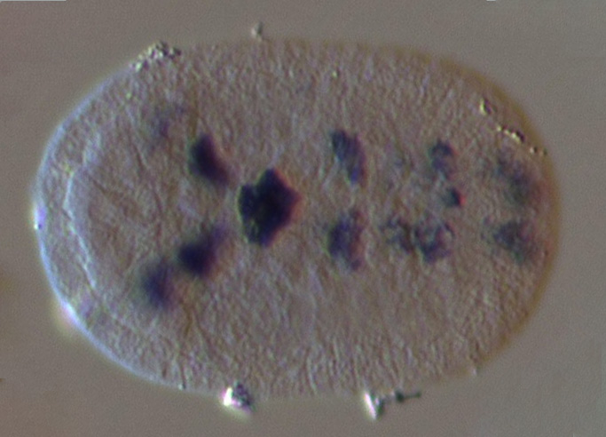
Vegetal view of a late gastrula stage embryo probed for neurogenin expression.

Original Annotation :
Neurogenin was expressed in A9.32 cell pair.

Aniseed Annotation :
According to the picture and to other data in Aniseed, there is also expression in the epidermis.

Stained molecule KH2012:KH.C6.129 (NEUROG1; NEUROG2; NEUROG3)
Stained region(s) A9.32 cell pair - dorsal midline anterior tail epidermis - lateral posterior tail epidermis -

C. robusta formely Ciona int. type A, Stage 12 (mid gastrula)

Read more…

Vegetal view of a late gastrula stage embryo probed for neurogenin expression, following the injection of Delta-like MO.

Original Annotation :
Neurogenin expression was lost in A9.32 cell pair (white arrow).

Stained molecule KH2012:KH.C6.129 (NEUROG1; NEUROG2; NEUROG3)
Stained region(s) dorsal midline anterior tail epidermis - lateral posterior tail epidermis -

C. robusta formely Ciona int. type A, Stage 12 (mid gastrula)

Read more…

Views of mid gastrula stage embryos showing reporter expression of the region upstream of neurogenin in A9.31 and A9.32 cells.

Stained molecule KH2012:KH.C6.129 (NEUROG1; NEUROG2; NEUROG3)
Stained region(s) A9.32 cell pair - posterior secondary muscle lineage (A9.31 line) -

C. robusta formely Ciona int. type A, Stage 13 (late gastrula)

Read more…

Dorsal view of a WT Ciona robusta late gastrula embryo probed for Neurogenin expression (magenta) and electroplated with Msx>LacZ (green). Neurogenin expression is observed in bipolar tail neurons (BTN) precursors and in neural plate border cells.
Original annotation : White arrowhead: Msx+/Neurog+ BTN progenitor. Black arrowhead: transient Neurog expression in BTN progenitor's sister cell (epidermal progenitor). Dashed line: midline. Scale bar, 25 μm.

Stained molecule KH2012:KH.C6.129 (NEUROG1; NEUROG2; NEUROG3)
Stained region(s) b9.36 cell pair - b line neural plate - posterior bipolar tail epidermal neurone precursors (b8.21 line) -

C. robusta formely Ciona int. type A, Stage 13 (late gastrula)

Read more…

Vegetal view of a late gastrula stage wild type embryo probed for Neurogenin expression.

Stained molecule KH2012:KH.C6.129 (NEUROG1; NEUROG2; NEUROG3)
Stained region(s) dorsal midline anterior tail epidermis - lateral posterior tail epidermis - neural plate -

C. robusta formely Ciona int. type A, Stage 13 (late gastrula)

Read more…

Conversion to Aniseed format of data from Yutaka Satou's gene expression profiles of transcription factors and signalling molecule ISH screen (Development. 2004 131:4047-58).

Original annotation :
Gene name :Neurogenin.
Expression pattern : nervous system.

Aniseed annotation : a staining has been detected in the neural plate but also in the A9.32 cell pair.

Stained molecule KH2012:KH.C6.129 (NEUROG1; NEUROG2; NEUROG3)
Stained region(s) dorsal midline anterior tail epidermis - lateral posterior tail epidermis - neural plate -

C. robusta formely Ciona int. type A, Stage 13 (late gastrula)

Read more…

Vegetal view of a late gastrula stage embryo probed for Onecut (HNF6) and Neurogenin expression (respectively labelled in red and green).
Original annotation : At late gastrula stage, Neurogenin expression continues to be detected in the A9.32 cells pair (white arrowheads), descendant of the A8.16, and begins to appear in two bilateral cells of the anterior neural plate, which will give rise to part of the sensory vesicle (green arrowheads). At this stage, Neuro- genin is also detected in the posterior part of the embryo at level of the neural tube and in the tail epidermal neuron precursors (blue arrowheads).
First image : Neurogenin ISH channel only.
Second image : Yellow color in the merged image represents co-expression territories.
Third image : Embryo used for imaging.

Stained molecule KH2012:KH.C6.129 (NEUROG1; NEUROG2; NEUROG3)
Stained region(s) A9.32 cell pair - a9.49 cell pair - a9.50 cell pair -

C. robusta formely Ciona int. type A, Stage 13 (late gastrula)

Read more…

Vegetal view of a late gastrula stage embryo probed for Neurogenin expression following egg micro-injection of a Nodal MO. Neurogenin was down-regulated in A-line nerve cord cells.

Aniseed annotation : weak staining can be detected in B-line muscle cells in the picture.

Stained molecule KH2012:KH.C6.129 (NEUROG1; NEUROG2; NEUROG3)
Stained region(s) primary muscle lineage -

C. robusta formely Ciona int. type A, Stage 13 (late gastrula)

Read more…

Vegetal view of a late gastrula stage embryo probed for Neurogenin expression following egg micro-injection of a SoxC MO. Neurogenin was ectopically expressed in medial part of A-line nerve cord cells.

Aniseed annotation : Weak staining can be detected in B-line muscle cells in the picture.

Stained molecule KH2012:KH.C6.129 (NEUROG1; NEUROG2; NEUROG3)
Stained region(s) A line neural plate - primary muscle lineage -

C. robusta formely Ciona int. type A, Stage 13 (late gastrula)

Read more…

Dorsal view of a WT Ciona robusta late gastrula embryo electroporated with Nodal b-line>H2B::Venus (red) and Neurog(Full lenght)>H2B::CFP (blue).
CFP staining is observed in b10.71, b10.72, and b8.21 lineage from b-line tail epidermis, .
Original annotation : Late gastrula embryo (stage 13) electroporated with full-length Neurog (blue) and Nodal b-line (red) reporter constructs. Reporter co-expression is seen in b9.36 descendants on either side of the neural plate. Neurog expression also marks tail-tip lineages of uncertain provenance, previously reported to be descended from b8.21. Scale bar, 25 μm

Stained molecule KH2012:KH.C6.129 (NEUROG1; NEUROG2; NEUROG3)
Stained region(s) dorsal midline anterior tail epidermis - posterior bipolar tail epidermal neurone precursors (b8.21 line) -

C. robusta formely Ciona int. type A, Stage 14 (early neurula)

Read more…

Neurula stage embryo probed for Neurogenin expression.

Stained molecule KH2012:KH.C6.129 (NEUROG1; NEUROG2; NEUROG3)
Stained region(s) neural plate -

C. robusta formely Ciona int. type A, Stage 14 (early neurula)

Read more…

Vegetal view of a neurula stage embryo probed for Neurogenin expression following egg micro-injection of a FoxD-a/b MO. Neurogenin is down-regulated in a-line neural cells.

Stained molecule KH2012:KH.C6.129 (NEUROG1; NEUROG2; NEUROG3)
Stained region(s) No expression -

C. robusta formely Ciona int. type A, Stage 15 (mid neurula)

Read more…

Conversion to Aniseed format of data from Yutaka Satou's gene expression profiles of transcription factors and signalling molecule ISH screen (Development. 2004 131:4047-58).

Original annotation :
Gene name :Neurogenin.
Expression pattern : nervous system

Stained molecule KH2012:KH.C6.129 (NEUROG1; NEUROG2; NEUROG3)
Stained region(s) neural plate -

C. robusta formely Ciona int. type A, Stage 15 (mid neurula)

Read more…

Vegetal view of a neurula probed for Onecut (HNF6) and Neurogenin expression (respectively labelled in green and red).
Original annotation : At neurula stage, Onecut is expressed in the tail neurons (blue arrowheads) and in the visceral ganglion precursors in the same cells also expressing Neurogenin (yellow arrowheads). At level of the anterior neural plate, Neurogenin shows a wide expression in four bilateral cell groups while Onecut is restricted to the central region, between the Neurogenin expressing cells.
First image : Neurogenin ISH channel only.
Second image : Yellow color in the merged image represents co-expression territories.
Third image : Embryo used for imaging.

Stained molecule KH2012:KH.C6.129 (NEUROG1; NEUROG2; NEUROG3)
Stained region(s) neural plate -

C. robusta formely Ciona int. type A, Stage 16 (late neurula)

Read more…

Dorsal view of a late neurula-stage embryo probed for Onecut (HNF6) and Neurogenin expression (respectively labelled in green and red).
Original annotation : At late neurula stage, the two genes are perfectly co- expressed in the visceral ganglion and caudal nerve cord precursors, while in the future sensory vesicle they still present different regions of expression and there are only few cells that co-express both genes that seem to represent the photoreceptor cell precursors (pink arrowheads).
First image : Neurogenin ISH channel only.
Second image : Yellow color in the merged image represents co-expression territories.
Third image : Embryo used for imaging.

Stained molecule KH2012:KH.C6.129 (NEUROG1; NEUROG2; NEUROG3)
Stained region(s) neural plate -

C. robusta formely Ciona int. type A, Stage 17 (initial tailbud I)

Read more…

Conversion to Aniseed format of Ghost database entry from Satou Y et al., Development 2001; 128:2893-2904. Original annotation: . Expression pattern: Mesenchyme Trunk lateral cells

Stained molecule KH2012:KH.C6.129 (NEUROG1; NEUROG2; NEUROG3)
Stained region(s) trunk lateral cells (A7.6 line) -

C. robusta formely Ciona int. type A, Stage 18 (initial tailbud II)

Read more…

Dorsal view of an early tailbud probed for Onecut (HNF6) and Neurogenin expression (respectively labelled in green and red).
Original annotation : At early tailbud stage, the merge images obtained by confocal microscopy demonstrates that these two genes are co-expressed in almost all the Onecut territories at level of the future sensory vesicle, visceral ganglion and caudal nerve cord, again with the exception of two small areas in the most anterior part of the nervous system corresponding to part of the future anterior sensory vesicle
First image : Neurogenin ISH channel only.
Second image : Yellow color in the merged image represents co-expression territories.
Third image : Embryo used for imaging.

Stained molecule KH2012:KH.C6.129 (NEUROG1; NEUROG2; NEUROG3)
Stained region(s) anterior sensory vesicle - posterior sensory vesicle - tail nerve cord - visceral ganglion -

C. robusta formely Ciona int. type A, Stage 19 (early tailbud I)

Read more…

Conversion to Aniseed format of Ghost database entry from Satou Y et al., Development 2001; 128:2893-2904. Original annotation: . Expression pattern: Mesenchyme Trunk lateral cells

Stained molecule KH2012:KH.C6.129 (NEUROG1; NEUROG2; NEUROG3)
Stained region(s) trunk lateral cells (A7.6 line) -

C. robusta formely Ciona int. type A, Stage 19 (early tailbud I)

Read more…

Conversion to Aniseed format of data from Yutaka Satou's gene expression profiles of transcription factors and signalling molecule ISH screen (Development. 2004 131:4047-58).

Original annotation :
Gene name :Neurogenin.
Expression pattern : nervous system, nerve cord.
Comment : 5 pairs of neural cells in the nerve cord, brain.

Aniseed annotation : a staining has been detected in the nervous system but also in the tail nerve cord.

Stained molecule KH2012:KH.C6.129 (NEUROG1; NEUROG2; NEUROG3)
Stained region(s) nervous system -

C. robusta formely Ciona int. type A, Stage 19 (early tailbud I)

Read more…

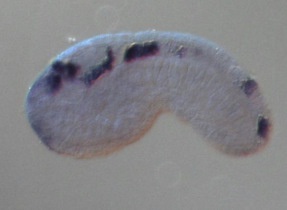
Original annotation : Ci-neurogenin expression at early tailbud, showing transcripts restricted to the central nervous system.

Aniseed annotation : according to the picture, it seems that expression is restricted to the brain and to the anterior part of the tail nerve cord.

Stained molecule KH2012:KH.C6.129 (NEUROG1; NEUROG2; NEUROG3)
Stained region(s) central nervous system -

C. robusta formely Ciona int. type A, Stage 19 (early tailbud I)

Read more…

Dorsal and lateral views of an early tailbud stage embryo, showing expression of Ci-NeuroD. Expression of Ci-NeuroD was revealed by whole-mount in situ hydridization.

Stained molecule KH2012:KH.C6.129 (NEUROG1; NEUROG2; NEUROG3)
Stained region(s) anterior sensory vesicle - posterior sensory vesicle - tail nerve cord -

C. robusta formely Ciona int. type A, Stage 19 (early tailbud I)

Read more…

Dorsal view of an early tailbud stage embryo probed for neurogenin expression.
Red and yellow arrowheads indicate boundaries between the neck and the visceral ganglion, and between the visceral ganglion and the caudal nerve cord, respectively.

Original Annotation :
Neurogenin was expressed in the visceral ganglion (A11.120, A11.119, A11.118, A11.117, A10.57 cell pairs).

Aniseed Annotation :
According to the picture, it seems that there is also expression in the posterior visceral ganglion.

Stained molecule KH2012:KH.C6.129 (NEUROG1; NEUROG2; NEUROG3)
Stained region(s) posterior sensory vesicle - visceral ganglion -

C. robusta formely Ciona int. type A, Stage 19 (early tailbud I)

Read more…

Dorsal view of an early tailbud stage embryo probed for neurogenin expression, following the injection of Cdx MO.
Red and yellow arrowheads indicate boundaries between the neck and the visceral ganglion, and between the visceral ganglion and the caudal nerve cord, respectively.

Original Annotation:
Neurogenin became ectopic in the nerve cord (A11.115 and A11.116 cell pair) (black arrow).

Aniseed Annotation:
It seems that there is also expression in the neck and in the posterior visceral ganglion.

Stained molecule KH2012:KH.C6.129 (NEUROG1; NEUROG2; NEUROG3)
Stained region(s) neck - posterior sensory vesicle - tail nerve cord - visceral ganglion -

C. robusta formely Ciona int. type A, Stage 19 (early tailbud I)

Read more…

Lateral view of an early tailbud stage embryo probed for neurogenin expression.

Stained molecule KH2012:KH.C6.129 (NEUROG1; NEUROG2; NEUROG3)
Stained region(s) anterior sensory vesicle - tail nerve cord - visceral ganglion -

C. robusta formely Ciona int. type A, Stage 19 (early tailbud I)

Read more…

Lateral view of an early tailbud stage embryo probed for neurogenin expression.

Original annotation:
Ectopic expression was observed (black arrow).

Stained molecule KH2012:KH.C6.129 (NEUROG1; NEUROG2; NEUROG3)
Stained region(s) anterior sensory vesicle - posterior tail epidermis - tail nerve cord - visceral ganglion -

C. robusta formely Ciona int. type A, Stage 19 (early tailbud I)

Read more…

Lateral view of the motor ganglion of a WT Ciona robusta early tailbud embryo probed for Ngn expression (red) and electroporated with FGF8/17/18>H2B:CFP reporter construct (blue nuclei).
Ngn is expressed in A11.120, A11.119, A11.118 and A11.117 motor neuron precursors.
Original comment:We observed initial expression in all four granddaughter cells of A9.30.
The A9.30 lineage (A11.120, A11.119, A11.118 and A11.117) is visualized using the FGF8/17/18 reporter construct (blue nuclei).

Stained molecule KH2012:KH.C6.129 (NEUROG1; NEUROG2; NEUROG3)
Stained region(s) visceral ganglion -

C. robusta formely Ciona int. type A, Stage 19 (early tailbud I)

Read more…

Views of early tailbud stage embryos showing reporter expression of the region upstream of neurogenin in nerve cord, muscle and mesenchyme.

Aniseed Annotation : According to the pictures it seems that in few embryos there also is staining (weak) in the sensory vesicle.

Stained molecule KH2012:KH.C6.129 (NEUROG1; NEUROG2; NEUROG3)
Stained region(s) posterior sensory vesicle - tail muscles - tail nerve cord -

C. robusta formely Ciona int. type A, Stage 20 (early tailbud II)

Read more…

Lateral and ventral views of a WT Ciona robusta early tailbud embryo probed for Neurogenin expression (green) and electroporated with Neurog b-line>unc76::mCherry (red). Neurogenin expression is observed in b11.141 but not in b11.142 cell pair
Original annotation : a, Lateral view of in situ hybridization (ISH) for Neurog (green) in embryo electroporated with Neurog b-line>H2B::mCherry (red) shows that Neurog expression is selectively maintained in only a subset of initially Neurog- expressing neural plate border cells. a′, In the b9.36 lineage, the anterior-most cell (b11.141, solid arrowhead) is always the sole one to express Neurog at this stage, and will go on to become the anterior BTN. Dashed arrowhead indicates b11.142, the sister cell of b11.141, which has downregulated Neurog relative to its sibling. b, b′, The identities of the cells in the tail tip (presumed b8.21-derived) lineages are unclear, but Neurog is similarly restricted (arrowheads) to a single cell on either side of the midline, which we interpret as the definitive posterior BTNs.
Scale bars 25 μm.

Stained molecule KH2012:KH.C6.129 (NEUROG1; NEUROG2; NEUROG3)
Stained region(s) anterior bipolar tail epidermal neurones (b8.18 line) - dorsal midline anterior tail epidermis - posterior bipolar tail epidermal neurones (b8.21 line) -

C. robusta formely Ciona int. type A, Stage 20 (early tailbud II)

Read more…

Zoomed view of the neural plate border of a WT Ciona robusta early tailbud embryo probed for neurogenin expression (green) and electroporated with Neurog b-line>H2B::mCherry (red).
Original annotation : Control embryo treated with DMSO vehicle, showing wild-type pattern of Neurog expression only in b11.141.
Scale bar 10 μm.

Stained molecule KH2012:KH.C6.129 (NEUROG1; NEUROG2; NEUROG3)
Stained region(s) anterior bipolar tail epidermal neurones (b8.18 line) -

C. robusta formely Ciona int. type A, Stage 20 (early tailbud II)

Read more…

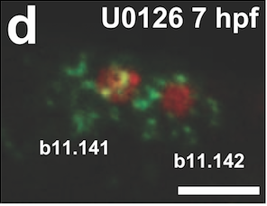
Zoomed view of the neural plate border of a Ciona robusta early tailbud embryo probed for neurogenin expression (green), electroporated with Neurog b-line>H2B::mCherry (red) and treated with the MEK inhibitor U0126 at 7 hours post fertilization.
Neurogenin expression is observed in both b11.141 and b 11.142 cell pairs. Original annotation : Neurog is expanded to b11.142 upon treatment with the MEK inhibitor U0126 at 7 h.p.f. This condition also results in specification of supernumerary BTNs, presumably due to expanded Neurog expression.
Scale bar 10 μm.

Stained molecule KH2012:KH.C6.129 (NEUROG1; NEUROG2; NEUROG3)
Stained region(s) anterior bipolar tail epidermal neurones (b8.18 line) - dorsal midline anterior tail epidermis -

C. robusta formely Ciona int. type A, Stage 20 (early tailbud II)

Read more…

Lateral view of WT Ciona robusta early tailbud embryo electroporated with Neurog b-line>unc-76::eGFP (green) counterstained with phalloidin (magenta).
eGFP is fused to UNC-76 tags to ensure even labelling of axons. GFP signalling is observed in bipolar tail neurons precursors(BTNs).
Original annotation : Embryos electroporated with Neurog b-line>unc-76::eGFP. BTN precursor (arrowhead) extending a lamellipodium. Scale bar, 25 μm.

Stained molecule KH2012:KH.C6.129 (NEUROG1; NEUROG2; NEUROG3)
Stained region(s) anterior bipolar tail epidermal neurones (b8.18 line) - posterior bipolar tail epidermal neurones (b8.21 line) -

C. robusta formely Ciona int. type A, Stage 20 (early tailbud II)

Read more…

Lateral view of a full embryo (left pannel) and the tail (right pannel) of a WT Ciona robusta early tailbud embryo co-electroporated with Neurog b-line>unc-76::VenusYFP (green) and Nodal b8.18>H2B::mCherry (magenta).
VenusYFP signalling is observed in the posterior dorsal midline tail epidermis including the bipolar tail neurons.
Original annotation : Embryos co-electroporated with Neurog b-line>unc-76::VenusYFP (green) and Nodal b8.18>H2B::mCherry (magenta). Right panels are enlarged views of the images on the left. Dashed lines indicate displacement from clonally related epidermal cells (indicated by colour-coded brackets). Scale bars, 25 μm.

Stained molecule KH2012:KH.C6.129 (NEUROG1; NEUROG2; NEUROG3)
Stained region(s) anterior bipolar tail epidermal neurones (b8.18 line) - dorsal midline posterior tail epidermis - posterior bipolar tail epidermal neurones (b8.21 line) -

C. robusta formely Ciona int. type A, Stage 20 (early tailbud II)

Read more…

Lateral and ventral views of a WT Ciona robusta early tailbud embryo probed for Neurogenin expression (green) and electroporated with Neurog b-line>H2B::mCherry (red). Neurog b line cis-regulatory sequence drives mCherry expression in the nuclei of b11.141, b11.142, b11.143 and b11.144 cell pairs. b11.141 is the anterior bipolar tail neuron (aBTN) precursor.
Boxed areas are enlarged in a' and b'. Scale bars 25 μm.

Stained molecule KH2012:KH.C6.129 (NEUROG1; NEUROG2; NEUROG3)
Stained region(s) anterior bipolar tail epidermal neurones (b8.18 line) - dorsal midline anterior tail epidermis -

C. robusta formely Ciona int. type A, Stage 21 (mid tailbud I)

Read more…

Conversion to Aniseed format of Ghost database entry from Satou Y et al., Development 2001; 128:2893-2904. Original annotation: . Expression pattern: Mesenchyme Trunk lateral cells

Stained molecule KH2012:KH.C6.129 (NEUROG1; NEUROG2; NEUROG3)
Stained region(s) trunk lateral cells (A7.6 line) -

C. robusta formely Ciona int. type A, Stage 21 (mid tailbud I)

Read more…

Conversion to Aniseed format of data from Yutaka Satou's gene expression profiles of transcription factors and signalling molecule ISH screen (Development. 2004 131:4047-58).

Original annotation :
Gene name :Neurogenin.
Expression pattern : nervous system, nerve cord.
Comment : 5 pairs of neural cells in the nerve cord, brain.

Aniseed annotation : a staining has been detected in the nervous system but also in the tail nerve cord and the visceral ganglion. A stianing has been detected in a cell in the tip of the tail epidermis.

Stained molecule KH2012:KH.C6.129 (NEUROG1; NEUROG2; NEUROG3)
Stained region(s) nervous system - tail epidermis -

C. robusta formely Ciona int. type A, Stage 21 (mid tailbud I)

Read more…

Mid tailbud stage embryo probed for neurogenin expression. No picture available in the article.

Original Annotation :
Expression of neurogenin in the visceral ganglion is localized in A12.240, A12.238, A13.474, A13.473, A12.239, A11.118, A11.117 cell pairs.

Stained molecule KH2012:KH.C6.129 (NEUROG1; NEUROG2; NEUROG3)
Stained region(s) visceral ganglion -

C. robusta formely Ciona int. type A, Stage 21 (mid tailbud I)

Read more…

Lateral view of the motor ganglion of a WT Ciona robusta mid tailbud embryo probed for Ngn (red) and HesB (green) expressions and electroporated with FGF8/17/18>H2B:CFP reporter construct (blue nuclei).
Original comment: At the mid-tailbud stage, Ngn expression (red) has refined to presumptive neurons A12.239, A13.474, A11.118 and A11.117. This alternates with HesB expression (green), in accordance with general models of lateral inhibition, except in A11.118/A11.117 pair (asterisks).
The A9.30 lineage is visualized using the FGF8/17/18 reporter construct (blue nuclei).

Stained molecule KH2012:KH.C6.129 (NEUROG1; NEUROG2; NEUROG3)
Stained region(s) visceral ganglion -

Lateral view of the motor ganglion of a WT Ciona robusta mid tailbud embryo probed for Ngn (red) and HesB (green) expressions and electroporated with FGF8/17/18>H2B:CFP reporter construct (blue nuclei).
Original comment: At the mid-tailbud stage, Ngn expression (red) has refined to presumptive neurons A12.239, A13.474, A11.118 and A11.117. This alternates with HesB expression (green), in accordance with general models of lateral inhibition, except in A11.118/A11.117 pair (asterisks).
The A9.30 lineage is visualized using the FGF8/17/18 reporter construct (blue nuclei).

Stained molecule KH2012:KH.C3.312 (HES1; HES4; HES6)
Stained region(s) visceral ganglion -

C. robusta formely Ciona int. type A, Stage 21 (mid tailbud I)

Read more…

Lateral view of a WT Ciona robusta mid tailbud embryo electroporated with Neurog b-line>unc-76::Venus .
Venus is fused to UNC-76 tags to ensure even labelling of axons. Venus signalling is observed in migrating bipolar tail neurons precursors (BTNs).
Original annotation :Migrating BTN precursors (arrowheads) labelled by the b-line-specific Neurog b-line>unc-76::Venus reporter construct (green). Scale bar, 25 μm.

Stained molecule KH2012:KH.C6.129 (NEUROG1; NEUROG2; NEUROG3)
Stained region(s) anterior bipolar tail epidermal neurones (b8.18 line) - dorsal midline anterior tail epidermis - dorsal midline posterior tail epidermis - posterior bipolar tail epidermal neurones (b8.21 line) -

C. robusta formely Ciona int. type A, Stage 21 (mid tailbud I)

Read more…

Lateral view of a WT Ciona robusta mid tailbud embryo electroporated with Neurog b-line>unc-76::eGFP (green) and counterstained with phalloidin (magenta).
GFP is fused to UNC-76 tags to ensure even labelling of axons. GFP signalling is observed in bipolar tail neuron (BTN) precursors of Ciona larvae.
Original annotation : Embryos electroporated with Neurog b-line>unc-76::eGFP. BTN precursors (anterior (aBTN) and posterior (pBTN)) delaminating. Scale bar, 25 μm.
Second picture: Enlarged view of aBTN. Top,UNC-76::eGFP; middle, phalloidin; bottom, merged; arrowhead, part of aBTN still in the epithelium. Scale bar, 10 μm.

Stained molecule KH2012:KH.C6.129 (NEUROG1; NEUROG2; NEUROG3)
Stained region(s) anterior bipolar tail epidermal neurones (b8.18 line) - dorsal midline posterior tail epidermis - posterior bipolar tail epidermal neurones (b8.21 line) -

C. robusta formely Ciona int. type A, Stage 21 (mid tailbud I)

Read more…

Lateral view of a full embryo (left pannel) and the tail (right pannel) of a WT Ciona robusta early tailbud embryo co-electroporated with Neurog b-line>unc-76::VenusYFP (green) and Nodal b8.18>H2B::mCherry (magenta).
VenusYFP signalling is observed in the posterior dorsal midline tail epidermis including the bipolar tail neurons.
Original annotation : Embryos co-electroporated with Neurog b-line>unc-76::VenusYFP (green) and Nodal b8.18>H2B::mCherry (magenta). Right panels are enlarged views of the images on the left. Dashed lines indicate displacement from clonally related epidermal cells (indicated by colour-coded brackets). Scale bars, 25 μm.

Stained molecule KH2012:KH.C6.129 (NEUROG1; NEUROG2; NEUROG3)
Stained region(s) anterior bipolar tail epidermal neurones (b8.18 line) - dorsal midline posterior tail epidermis - posterior bipolar tail epidermal neurones (b8.21 line) -

C. robusta formely Ciona int. type A, Stage 22 (mid tailbud II)

Read more…

Dorso-latteral view of a tailbud probed for Onecut (HNF6) and Neurogenin expression (respectively labelled in red and green).
Original annotation : A middle tailbud stage it is possible to observe a full co-localization of the two genes not only in the visceral ganglion and dorsal nerve cord, but also in the anterior sensory vesicle precursors, where in the previous stages there was only Onecut (yellow arrowheads).
First image : Neurogenin ISH channel only.
Second image : Yellow color in the merged image represents co-expression territories, edge of the embryo is highlighted with a thin white line.

Stained molecule KH2012:KH.C6.129 (NEUROG1; NEUROG2; NEUROG3)
Stained region(s) anterior sensory vesicle - posterior sensory vesicle - tail nerve cord - visceral ganglion -

C. robusta formely Ciona int. type A, Stage 22 (mid tailbud II)

Read more…

Lateral view of a WT Ciona robusta mid tailbud embryo electroporated with Neurog b-line>H2B::Venus.
Venus signalling is observed in dorsal b-line, and tip of the tail epidermis
Original annotation : This Neurog cis-regulatory element drives reporter gene expression in the caudal neural plate border region.

Stained molecule KH2012:KH.C6.129 (NEUROG1; NEUROG2; NEUROG3)
Stained region(s) dorsal midline posterior tail epidermis - dorso lateral posterior tail epidermis -

C. robusta formely Ciona int. type A, Stage 22 (mid tailbud II)

Read more…

Lateral view of a WT Ciona robusta mid tailbud embryo electroporated with Neurog b-line minimal>H2B::mCherry.
mCherry signalling is observed in some cells of the posterior dorsal tail epidermis.

Stained molecule KH2012:KH.C6.129 (NEUROG1; NEUROG2; NEUROG3)
Stained region(s) dorsal midline posterior tail epidermis -

C. robusta formely Ciona int. type A, Stage 22 (mid tailbud II)

Read more…

Lateral view of the tail of a WT Ciona robusta mid tailbud embryo co-electroporated with Mrf>unc-76::eGFP (green) and Neurog b-line>unc76::mCherry (magenta), and counterstained with phalloidin.
mCherry signalling is observed in the bipolar tail neurons.
Original annotation : Embryo with paraxial mesoderm labelled by Mrf>unc-76::eGFP (green), BTN labelled by Neurog b-line>unc- 76::mCherry (magenta) and phalloidin counterstain. Scale bar, 25 μm. Right, cross-sectioned 3D image of same embryo. Only the right side of the embryo was transfected.

Stained molecule KH2012:KH.C6.129 (NEUROG1; NEUROG2; NEUROG3)
Stained region(s) anterior bipolar tail epidermal neurones (b8.18 line) - dorsal midline posterior tail epidermis - posterior bipolar tail epidermal neurones (b8.21 line) -

C. robusta formely Ciona int. type A, Stage 22 (mid tailbud II)

Read more…

3D slice of the tail of a WT Ciona robusta mid tailbud embryo electroporated with Neurog b-line>unc76::mCherry and counterstained with phalloidin.
mCherry signalling is observed in the bipolar tail neurons (BTNs).
Original annotation : 3D slice of embryo showing BTNs (arrowheads) outside neural tube (dotted outline). Only the right side of the embryo was transfected.

Stained molecule KH2012:KH.C6.129 (NEUROG1; NEUROG2; NEUROG3)
Stained region(s) anterior bipolar tail epidermal neurones (b8.18 line) - posterior bipolar tail epidermal neurones (b8.21 line) -

C. robusta formely Ciona int. type A, Stage 22 (mid tailbud II)

Read more…

Lateral view of a full embryo (left pannel) and the tail (right pannel) of a WT Ciona robusta mid tailbud embryo co-electroporated with Neurog b-line>unc-76::VenusYFP (green) and Nodal b8.18>H2B::mCherry (magenta).
VenusYFP signalling is observed in the posterior dorsal midline tail epidermis and in the bipolar tail neurons (pBTN and aBTN, white and pink arrowheads respectively).
Original annotation : Dashed lines indicate displacement from clonally related epidermal cells (indicated by colour-coded brackets). Scale bars, 25 μm.

Stained molecule KH2012:KH.C6.129 (NEUROG1; NEUROG2; NEUROG3)
Stained region(s) anterior bipolar tail epidermal neurones (b8.18 line) - dorsal midline posterior tail epidermis - posterior bipolar tail epidermal neurones (b8.21 line) -

C. robusta formely Ciona int. type A, Stage 22 (mid tailbud II)

Read more…

Lateral view of a WT Ciona robusta mid tailbud embryo electroporated with Neurog b-line>unc-76::VenusYFP (green), treated with DMSO at 5.5hpf (control) and counterstained with DAPI (blue)
VenusYFP is fused to UNC-76 tags to ensure even labelling of axons.
VenusYFP signalling is observed in the posterior dorsal midline tail epidermis, in the bipolar tail neurons (BTNs), and in the mesenchyme.

Stained molecule KH2012:KH.C6.129 (NEUROG1; NEUROG2; NEUROG3)
Stained region(s) anterior bipolar tail epidermal neurones (b8.18 line) - dorsal midline posterior tail epidermis - mesenchyme (B8.5 & B7.7 lines) - posterior bipolar tail epidermal neurones (b8.21 line) -

C. robusta formely Ciona int. type A, Stage 22 (mid tailbud II)

Read more…

Lateral view of a Ciona robusta mid tailbud embryo electroporated with Neurog b-line>unc-76::VenusYFP (green), treated with U0126 at 5.5hpf and counterstained with DAPI (blue)
VenusYFP is fused to UNC-76 tags to ensure even labelling of axons.
No VenusYFP signalling is observed.
Original annotation : Neurog expression was abolished in 43 of 50 embryos treated with 10 μM MEK inhibitor U0126 at 5.5 hours post-fertilization (h.p.f.)

Stained molecule KH2012:KH.C6.129 (NEUROG1; NEUROG2; NEUROG3)
Stained region(s) No expression -

C. robusta formely Ciona int. type A, Stage 22 (mid tailbud II)

Read more…

Lateral view of a Ciona robusta mid tailbud embryo electroporated with Neurog b-line>unc-76::VenusYFP (green), treated with U0126 at 7.5hpf and counterstained with DAPI (blue)
VenusYFP is fused to UNC-76 tags to ensure even labelling of axons.
VenusYFP signalling is observed in the posterior dorsal midline of the tail epidermis and in supernumerary bippolar tail neurons (BTNs).
Original annotation : Supernumerary BTNs were specified in 28 of 50 embryos treated with 10 μM U0126 at 7 h.p.f

Stained molecule KH2012:KH.C6.129 (NEUROG1; NEUROG2; NEUROG3)
Stained region(s) anterior bipolar tail epidermal neurones (b8.18 line) - dorsal midline posterior tail epidermis - posterior bipolar tail epidermal neurones (b8.21 line) -

C. robusta formely Ciona int. type A, Stage 22 (mid tailbud II)

Read more…

Lateral view of the posterior dorsal tail midline of a WT Ciona robusta mid tailbud embryo co-electroporated with Neurog b-line>unc-76::VenusYFP (green) and Neurog b-line>H2B::mCherry (magenta), treated with DMSO at 7.5hpf (control experiment). VenusYFP is fused to UNC-76 tags to ensure even labelling of axons. VenusYFP signalling is observed in the bipolar tail neurons (BTNs).
Original annotation : Two BTN precursors, labelled by Neurog b-line>unc-76::VenusYFP (green) and Neurog b-line>H2B::mCherry (magenta), migrating on one side of a DMSO-treated embryo.

Stained molecule KH2012:KH.C6.129 (NEUROG1; NEUROG2; NEUROG3)
Stained region(s) anterior bipolar tail epidermal neurones (b8.18 line) - posterior bipolar tail epidermal neurones (b8.21 line) -

C. robusta formely Ciona int. type A, Stage 22 (mid tailbud II)

Read more…

Lateral view of the posterior dorsal tail midline of a Ciona robusta mid tailbud embryo co-electroporated with Neurog b-line>unc-76::VenusYFP (green) and Neurog b-line>H2B::mCherry (magenta), treated with U0126 MEK inhibitor at 7.5hpf.
VenusYFP is fused to UNC-76 tags to ensure even labelling of axons.
VenusYFP signalling is observed in an expending chain of four bipolar tail neurons (BTNs).

Stained molecule KH2012:KH.C6.129 (NEUROG1; NEUROG2; NEUROG3)
Stained region(s) anterior bipolar tail epidermal neurones (b8.18 line) - dorsal midline posterior tail epidermis - posterior bipolar tail epidermal neurones (b8.21 line) -

C. robusta formely Ciona int. type A, Stage 22 (mid tailbud II)

Read more…

Lateral view of the posterior dorsal tail midline of a WT Ciona robusta mid tailbud embryo co-electroporated with Neurog b-line>unc-76::VenusYFP (green) and Neurog b-line>H2B::mCherry (magenta), treated with DMSO at 7.5hpf (control experiment). mCherry signalling is observed in the bipolar tail neurons (BTNs).
Original annotation : Two BTN precursors, labelled by Neurog b-line>unc-76::VenusYFP (green) and Neurog b-line>H2B::mCherry (magenta), migrating on one side of a DMSO-treated embryo.

Stained molecule KH2012:KH.C6.129 (NEUROG1; NEUROG2; NEUROG3)
Stained region(s) anterior bipolar tail epidermal neurones (b8.18 line) - posterior bipolar tail epidermal neurones (b8.21 line) -

C. robusta formely Ciona int. type A, Stage 22 (mid tailbud II)

Read more…

Lateral view of the posterior dorsal tail midline of a Ciona robusta mid tailbud embryo co-electroporated with Neurog b-line>unc-76::VenusYFP (green) and Neurog b-line>H2B::mCherry (magenta), treated with U0126 MEK inhibitor at 7.5hpf.
mCherry signaling is observed in an expending chain of four bipolar tail neurons (BTNs).

Stained molecule KH2012:KH.C6.129 (NEUROG1; NEUROG2; NEUROG3)
Stained region(s) anterior bipolar tail epidermal neurones (b8.18 line) - dorsal midline posterior tail epidermis - posterior bipolar tail epidermal neurones (b8.21 line) -

C. robusta formely Ciona int. type A, Stage 22 (mid tailbud II)

Read more…

Lateral view of the posterior dorsal tail of a WT Ciona robusta mid tailbud embryo electroporated with Neurog b-line>unc-76::mCherry (magenta) and probed for Cadherin.b expression (green).
mCherry signalling is observed in the bipolar tail neuron precursors.

Stained molecule KH2012:KH.C6.129 (NEUROG1; NEUROG2; NEUROG3)
Stained region(s) anterior bipolar tail epidermal neurones (b8.18 line) - posterior bipolar tail epidermal neurones (b8.21 line) -

C. robusta formely Ciona int. type A, Stage 22 (mid tailbud II)

Read more…

Lateral view of the posterior dorsal tail of a WT Ciona robusta mid tailbud embryo electroporated with Neurog b-line>unc-76::mCherry (magenta) and probed for Cadherin.b expression (green).
mCherry signalling is observed in the bipolar tail neuron precursors.

Stained molecule KH2012:KH.C6.129 (NEUROG1; NEUROG2; NEUROG3)
Stained region(s) anterior bipolar tail epidermal neurones (b8.18 line) - dorsal midline posterior tail epidermis - posterior bipolar tail epidermal neurones (b8.21 line) -

C. robusta formely Ciona int. type A, Stage 22 (mid tailbud II)

Read more…

Lateral view of a WTCiona robusta tailbud embryo (11.5 hpf) co-electroporated with Neurog b-line>unc76::VENUS7FP (red), Neurog b-line>Galnt7ΔC::CFP (green) and Neurog b-line>H2B::mCherry (blue). Neurog b-line cis-regulatory element drives reporter expression in b-line epidermis including anterior and posterior bipolar tail neurons (aBTN and pBTN).
unc76 tag is used to label axons.
H2B tag is used to label nuclei.
Galnt7ΔC tag is used to label Golgi.
Original annotation : Targeted localization of CFP by the Galnt7 N-terminal signal sequence reveals polarized subcellular distribution of Golgi apparatus on posterior side of BTN nuclei as migration and proximal process extend in an anterior direction. This is distinct from the apical (dorsal) location of the Golgi apparatus in epidermal cells.
Scale bar 50 μm.

Stained molecule KH2012:KH.C6.129 (NEUROG1; NEUROG2; NEUROG3)
Stained region(s) anterior bipolar tail epidermal neurones (b8.18 line) - anterior tail epidermis - dorsal midline anterior tail epidermis - posterior bipolar tail epidermal neurones (b8.21 line) -

Lateral view of the tail of a WT Ciona robusta tailbud embryo (11.5 hpf) co-electroporated with Neurog b-line>unc76::VENUS7FP (red), Neurog b-line>Galnt7ΔC::CFP (green) and Neurog b-line>H2B::mCherry (blue). Neurog b-line cis-regulatory element drives reporter expression in b-line epidermis including anterior and posterior bipolar tail neurons (aBTN and pBTN).
unc76 tag is used to label axons.
H2B tag is used to label nuclei.
Galnt7ΔC tag is used to label Golgi apparatus.
Original annotation : Targeted localization of CFP by the Galnt7 N-terminal signal sequence reveals polarized subcellular distribution of Golgi apparatus on posterior side of BTN nuclei as migration and proximal process extend in an anterior direction. This is distinct from the apical (dorsal) location of the Golgi apparatus in epidermal cells.
Scale bar 50 μm.

Stained molecule KH2012:KH.C6.129 (NEUROG1; NEUROG2; NEUROG3)
Stained region(s) anterior bipolar tail epidermal neurones (b8.18 line) - anterior tail epidermis - dorsal midline anterior tail epidermis - posterior bipolar tail epidermal neurones (b8.21 line) -

C. robusta formely Ciona int. type A, Stage 22 (mid tailbud II)

Read more…

Lateral view of the tail of a WT Ciona robusta tailbud embryo (11.5 hpf) co-electroporated with Neurog b-line>unc76::VENUS7FP (red), Neurog b-line>Galnt7ΔC::CFP (green) and Neurog b-line>H2B::mCherry (blue). Neurog b-line cis-regulatory element drives reporter expression in b-line epidermis including anterior and posterior bipolar tail neurons (aBTN and pBTN).
unc76 tag is used to label axons.
H2B tag is used to label nuclei.
Galnt7ΔC tag is used to label Golgi apparatus.
Original annotation : Targeted localization of CFP by the Galnt7 N-terminal signal sequence reveals polarized subcellular distribution of Golgi apparatus on posterior side of BTN nuclei as migration and proximal process extend in an anterior direction. This is distinct from the apical (dorsal) location of the Golgi apparatus in epidermal cells.
Scale bar 50 μm.

Stained molecule KH2012:KH.C6.129 (NEUROG1; NEUROG2; NEUROG3)
Stained region(s) anterior bipolar tail epidermal neurones (b8.18 line) - dorsal midline anterior tail epidermis - posterior bipolar tail epidermal neurones (b8.21 line) -

C. robusta formely Ciona int. type A, Stage 22 (mid tailbud II)

Read more…

Posterior region of the tail of a mid tailbud-stage Ciona robusta embryo, coelectroporated with Islet>unc-76::GFP (green) and Ngn>mCherry (red) reporter constructs. The second image is the un-annotated HD picture.
Original annotation: Neurogenin (Ngn) cis-regulatory sequence drives mCherry expression in A- and b- neural plate border cells, which gives rise to ependymal cells (open arrowheads) and bipolar neurons (solid arrowheads) of the posterior tail nerve cord, as well as secondary tail muscle cells (“m”). The Islet driver only drives expression in bipolar tail neurons.

Stained molecule KH2012:KH.L152.2 (ISL1; ISL2; LHX9)
Stained region(s) posterior bipolar tail epidermal neurones (b8.21 line) - posterior bipolar tail epidermal neurones (b8.25 line) -

Posterior region of the tail of a mid tailbud-stage Ciona robusta embryo, coelectroporated with Islet>unc-76::GFP (green) and Ngn>mCherry (red) reporter constructs. The second image is the un-annotated HD picture.
Original annotation: Neurogenin (Ngn) cis-regulatory sequence drives mCherry expression in A- and b- neural plate border cells, which gives rise to ependymal cells (open arrowheads) and bipolar neurons (solid arrowheads) of the posterior tail nerve cord, as well as secondary tail muscle cells (“m”). The Islet driver only drives expression in bipolar tail neurons.

Stained molecule KH2012:KH.C6.129 (NEUROG1; NEUROG2; NEUROG3)
Stained region(s) posterior bipolar tail epidermal neurones (b8.21 line) - posterior bipolar tail epidermal neurones (b8.25 line) - secondary muscle lineage - TNC ependymal precursors -

C. robusta formely Ciona int. type A, Stage 23 (late tailbud I)

Read more…

Conversion to Aniseed format of Ghost database entry from Satou Y et al., Development 2001; 128:2893-2904. Original annotation: . Expression pattern: Mesenchyme Trunk lateral cells

Stained molecule KH2012:KH.C6.129 (NEUROG1; NEUROG2; NEUROG3)
Stained region(s) trunk lateral cells (A7.6 line) -

C. robusta formely Ciona int. type A, Stage 23 (late tailbud I)

Read more…

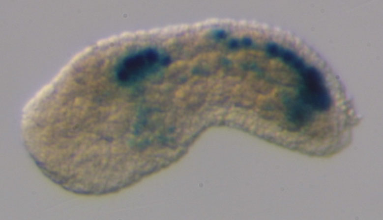
Original annotation : Ci-neurogenin expression at late tailbud, showing transcripts restricted to the central nervous system.

Aniseed annotation : according to the picture, expression seems to be restricted to the sensory vesicle and weakly to the visceral ganglion.

Stained molecule KH2012:KH.C6.129 (NEUROG1; NEUROG2; NEUROG3)
Stained region(s) anterior sensory vesicle - posterior sensory vesicle - visceral ganglion -

C. robusta formely Ciona int. type A, Stage 23 (late tailbud I)

Read more…

Lateral view of a late tailbud stage embryo, showing expression of Ci-NeuroD. Expression of Ci-NeuroD was revealed by whole-mount in situ hydridization.

Stained molecule KH2012:KH.C6.129 (NEUROG1; NEUROG2; NEUROG3)
Stained region(s) anterior sensory vesicle - posterior sensory vesicle -

C. robusta formely Ciona int. type A, Stage 23 (late tailbud I)

Read more…

Lateral view of the posterior tail of a Ciona robusta late tailbud embryo co-electroporated with Asic>unc76::eGFP (green) and Neurog b-line>nls::LacZ (red).
LacZ signalling is observed in the dorsal posterior midline tail epidermis, including the bipolar tail neurons (BTNs).

Stained molecule KH2012:KH.C6.129 (NEUROG1; NEUROG2; NEUROG3)
Stained region(s) anterior bipolar tail epidermal neurones (b8.18 line) - dorsal midline posterior tail epidermis - posterior bipolar tail epidermal neurones (b8.21 line) -

C. robusta formely Ciona int. type A, Stage 23 (late tailbud I)

Read more…

Lateral view of the posterior tail of a Ciona robusta late tailbud embryo co-electroporated with Asic>unc76::eGFP (green) and Neurog b-line>nls::LacZ (red), in which Neurogenin was overexpressed using Neurog b-line driver sequence.
LacZ signalling is observed in posterior dorsal tail epidermis, including supplementary bipolar tail neurons (BTNs).
Original annotation : Overexpression of Neurog induced ectopic migratory Asic+ BTN precursors.

Stained molecule KH2012:KH.C6.129 (NEUROG1; NEUROG2; NEUROG3)
Stained region(s) anterior bipolar tail epidermal neurones (b8.18 line) - dorsal midline posterior tail epidermis - dorso lateral posterior tail epidermis - lateral posterior tail epidermis - posterior bipolar tail epidermal neurones (b8.21 line) -

C. robusta formely Ciona int. type A, Stage 23 (late tailbud I)

Read more…

Lateral view of the posterior tail of a Ciona robusta late tailbud embryo co-electroporated with Asic>unc76::eGFP (green) and Neurog b-line>nls::LacZ (red), in which Neurogenin dominant repressor form was overexpressed using Neurog b-line driver sequence.
LacZ signalling is observed in posterior dorsal tail epidermis.
Original annotation : Overexpression of a dominant repressor form of Neurog (Neurog::WRPW) abolishes BTNs in 97 of 100 embryos.

Stained molecule KH2012:KH.C6.129 (NEUROG1; NEUROG2; NEUROG3)
Stained region(s) anterior bipolar tail epidermal neurones (b8.18 line) - dorsal midline posterior tail epidermis - dorso lateral posterior tail epidermis - posterior bipolar tail epidermal neurones (b8.21 line) -

C. robusta formely Ciona int. type A, Stage 23 (late tailbud I)

Read more…

Lateral view of a WT Ciona robusta late tailbud embryo co-electroporated with Neurog b-line>unc76::GFP (green) and Msx>H2B::mCherry (magenta nuclei). Msx cis-regulatory region drives mCherry expression in the neural tube.
Scale bar 50 μm

Stained molecule KH2012:KH.C2.957 (DLX1; MSX1; MSX2)
Stained region(s) tail nerve cord -

Lateral view of a WT Ciona robusta late tailbud embryo co-electroporated with Neurog b-line>unc76::GFP (green) and Msx>H2B::mCherry (magenta nuclei). Neurog b-line cis-regulatory region drives GFP expression in the bipolar tail neurons precursors
Scale bar 50 μm

Stained molecule KH2012:KH.C6.129 (NEUROG1; NEUROG2; NEUROG3)
Stained region(s) anterior bipolar tail epidermal neurones (b8.18 line) - dorsal midline posterior tail epidermis - posterior bipolar tail epidermal neurones (b8.21 line) -

C. robusta formely Ciona int. type A, Stage 23 (late tailbud I)

Read more…

Lateral view of a Ciona robusta late tailbud embryo co-electroporated with Neurog b-line>unc-76::GFP (green) and Msx>H2B::mCherry (magenta nuclei) and counterstained with phalloidin (magenta) and Msx>Su(H)-DBM, which encodes a DNA-binding mutant form of the Notch co-activator Rbpj. Msx cis-regulatory region drives mCherry expression in the neural tube
Original annotation : No discernable difference in Neurog activation or bipolar tail neuron specification was observed between control and Su(H)-DBM conditions (1 of 32 versus 2 of 42 embryos showing ectopic Neurog+ BTNs, respectively).
Scale bars 25 μm.

Stained molecule KH2012:KH.C2.957 (DLX1; MSX1; MSX2)
Stained region(s) tail nerve cord -

Lateral view of a Ciona robusta late tailbud embryo co-electroporated with Neurog b-line>unc-76::GFP (green) and Msx>H2B::mCherry (magenta nuclei) and counterstained with phalloidin (magenta) and Msx>Su(H)-DBM, which encodes a DNA-binding mutant form of the Notch co-activator Rbpj. Neurog b-line cis-regulatory element drives GFP expression in the bipolar tail neuron precursors.
Original annotation : No discernable difference in Neurog activation or bipolar tail neuron specification was observed between control and Su(H)-DBM conditions (1 of 32 versus 2 of 42 embryos showing ectopic Neurog+ BTNs, respectively).
Scale bars 25 μm.

Stained molecule KH2012:KH.C6.129 (NEUROG1; NEUROG2; NEUROG3)
Stained region(s) anterior bipolar tail epidermal neurones (b8.18 line) - dorsal midline anterior tail epidermis - posterior bipolar tail epidermal neurones (b8.21 line) -

C. robusta formely Ciona int. type A, Stage 23 (late tailbud I)

Read more…

Lateral view of a Ciona robusta late tailbud embryo co-electroporated with Neurog b-line>H2B::mCherry (magenta nuclei) and Asic>unc76::eGFP (green). Neurog b-line cis-regulatory element drives mCherry expression in the b-line tail epidermis including the bipolar tail neurons.
Scale bars 25 μm.

Stained molecule KH2012:KH.C6.129 (NEUROG1; NEUROG2; NEUROG3)
Stained region(s) anterior bipolar tail epidermal neurones (b8.18 line) - dorsal midline anterior tail epidermis - posterior bipolar tail epidermal neurones (b8.21 line) -

Lateral view of a Ciona robusta late tailbud embryo co-electroporated with Neurog b-line>H2B::mCherry (magenta nuclei) and Asic>unc76::eGFP (green). eGFP signalling is observed in the bipolar tail neurons (BTNs).
Scale bars 25 μm.

Stained molecule KH2012:KH.C1.215 (ASIC1; ASIC2; ASIC3)
Stained region(s) anterior bipolar tail epidermal neurones (b8.18 line) - posterior bipolar tail epidermal neurones (b8.21 line) -

C. robusta formely Ciona int. type A, Stage 23 (late tailbud I)

Read more…

Lateral view of a Ciona robusta late tailbud embryo co-electroporated with Neurog b-line>H2B::mCherry (magenta nuclei), Asic>unc76::eGFP (green) and Neurog b-line>Su(H)-DBM, which encodes a DNA-binding mutant form of the Notch co-activator Rbpj. Neurog b-line cis-regulatory element drives mCherry expression in the b-line tail epidermis including the bipolar tail neuron precursors.
Original annotation : Late overexpression of Su(H)-DBM using the Neurog b-line driver did not alter BTN (bipolar tail neuron) specification/differentiation, as monitored by Asic>unc- 76::eGFP reporter expression (0 of 50 control versus 0 of 50 Su(H)-DBM embryos showed ectopic Asic+ BTNs). .
Scale bars 25 μm.

Stained molecule KH2012:KH.C6.129 (NEUROG1; NEUROG2; NEUROG3)
Stained region(s) anterior bipolar tail epidermal neurones (b8.18 line) - anterior tail epidermis - posterior bipolar tail epidermal neurones (b8.21 line) -

Lateral view of a Ciona robusta late tailbud embryo co-electroporated with Neurog b-line>H2B::mCherry (magenta nuclei), Asic>unc76::eGFP (green) and Neurog b-line>Su(H)-DBM, which encodes a DNA-binding mutant form of the Notch co-activator Rbpj. Asic cis-regulatory element drives eGFP expression in the bipolar tail neurons (BTN).
Original annotation : Late overexpression of Su(H)-DBM using the Neurog b-line driver did not alter BTN specification/differentiation, as monitored by Asic>unc- 76::eGFP reporter expression (0 of 50 control versus 0 of 50 Su(H)-DBM embryos showed ectopic Asic+ BTNs). .
Scale bars 25 μm.

Stained molecule KH2012:KH.C1.215 (ASIC1; ASIC2; ASIC3)
Stained region(s) anterior bipolar tail epidermal neurones (b8.18 line) - posterior bipolar tail epidermal neurones (b8.21 line) -

C. robusta formely Ciona int. type A, Stage 23 (late tailbud I)

Read more…

Lateral view of the bipolar tail neurons (BTN) of a WT Ciona robusta tailbud embryo (12.5 hpf) co-electroporated with Neurog b-line>unc76::VENUSYFP (red), Neurog b-line>Galnt7ΔC::CFP (green) and Neurog b-line>H2B::mCherry (blue). Neurog b-line cis-regulatory element drives reporter expression in the anterior and posterior bipolar tail neurons (aBTN and pBTN).
unc76 tag is used to label axons.
H2B tag is used to label nuclei.
Galnt7ΔC tag is used to label Golgi apparatus.
Original annotation : Embryo at 12.5 h.p.f. (18 °C) showing 180° inversion of Golgi apparatus localization to the anterior side of the nucleus (compared to its localization on posterior side of BTN nuclei at 11.5 hpf), immediately preceding distal process extension.
Scale bar 50 μm.

Stained molecule KH2012:KH.C6.129 (NEUROG1; NEUROG2; NEUROG3)
Stained region(s) anterior bipolar tail epidermal neurones (b8.18 line) - posterior bipolar tail epidermal neurones (b8.21 line) -

C. robusta formely Ciona int. type A, Stage 23 (late tailbud I)

Read more…

Lateral view of the bipolar tail neurons (BTN) of a WT Ciona robusta tailbud embryo (12.5 hpf) co-electroporated with Neurog b-line>unc76::VENUSYFP (red), Neurog b-line>Galnt7ΔC::CFP (green) and Neurog b-line>H2B::mCherry (blue). Neurog b-line cis-regulatory element drives reporter expression in the anterior and posterior bipolar tail neurons (aBTN and pBTN).
unc76 tag is used to label axons.
H2B tag is used to label nuclei.
Galnt7ΔC tag is used to label Golgi apparatus.
Original annotation : Embryo at 12.5 h.p.f. (18 °C) showing 180° inversion of Golgi apparatus localization to the anterior side of the nucleus (compared to its localization on posterior side of BTN nuclei at 11.5 hpf), immediately preceding distal process extension.
Scale bar 50 μm.

Stained molecule KH2012:KH.C6.129 (NEUROG1; NEUROG2; NEUROG3)
Stained region(s) anterior bipolar tail epidermal neurones (b8.18 line) - posterior bipolar tail epidermal neurones (b8.21 line) -

C. robusta formely Ciona int. type A, Stage 25 (late tailbud III)

Read more…

Dorso-latteral view of a late tailbud probed for Onecut (HNF6) and Neurogenin expression (respectively labelled in red and green).
Original annotation : At larva stage, it is possible to observe an almost complete co-localization of the two genes in the Onecut territories (ie. sensory vesicle, visceral ganglion and part of the tail nerve chord).
First image : Neurogenin ISH channel only.
Second image : Yellow color in the merged image represents co-expression territories, edge of the embryo is highlighted with a thin white line.

Stained molecule KH2012:KH.C6.129 (NEUROG1; NEUROG2; NEUROG3)
Stained region(s) anterior sensory vesicle - posterior sensory vesicle - tail nerve cord - visceral ganglion -

C. robusta formely Ciona int. type A, Stage 26 (hatching larva)

Read more…

Latteral view of an early larva probed for Neurogenin and Arrestin expression (respectively labelled in red and green). Onecut signal is detected in the sensory vesicle and inthe visceral ganglion.
Original annotation : At larva stage, it is possible to observe an almost complete co-localization of the two genes in the Onecut territories.
First image : Neurogenin ISH channel only.
Second image : Yellow color in the merged image represents co-expression territories, edge of the embryo is highlighted with a thin white line.

Stained molecule KH2012:KH.C6.129 (NEUROG1; NEUROG2; NEUROG3)
Stained region(s) anterior sensory vesicle - posterior sensory vesicle - visceral ganglion -

C. robusta formely Ciona int. type A, Stage 26 (hatching larva)

Read more…

Views of larvae showing reporter expression of the region upstream of neurogenin in nerve cord, muscle and mesenchyme.

Aniseed Annotation : According to the second picture, in few embryos anterior sensory vesicle staining could be detected.

Stained molecule KH2012:KH.C6.129 (NEUROG1; NEUROG2; NEUROG3)
Stained region(s) tail muscles - tail nerve cord -

C. robusta formely Ciona int. type A, Stage 26 (hatching larva)

Read more…

Lateral view of the head of a WT Ciona robusta larva co-electroporated with wild-type (red) and mutated minimal (green) Dmbx reporter constructs.
Mutation of the predicted Pax3/7 site abolishes reporter activity (GFP expression seen in only 1/100 embryos).

Stained molecule KH2012:KH.C1.1212 (DMBX1; DRGX; PITX3)
Stained region(s) No expression -

Lateral view of the head of a WT Ciona robusta larva co-electroporated with wild-type (red) and mutated (green) minimal Dmbx reporter constructs.
WT Dmbx enhancer is active in A12.239 neuron pair.

Stained molecule KH2012:KH.C6.129 (NEUROG1; NEUROG2; NEUROG3)
Stained region(s) motor neurones -

C. robusta formely Ciona int. type A, Stage 48 (young adult)

Read more…

Left side view of a tadpole stage wild type embryo probed for Ci-neurogenin expression. Expression was detected in the ectoderm atria.

Stained molecule KH2012:KH.C6.129 (NEUROG1; NEUROG2; NEUROG3)
Stained region(s)