Login
Help

GENE CARD

Submit your Data

  1. EST 'FF899862.1'

EST count

27 results

Library Name Percentage Expression
Ciona intestinalis whole animal Ciona intestinalis whole animal 0 clone(s) / 0
Nori Satoh unpublished cDNA library Nori Satoh unpublished cDNA library 1 clone(s) / 5,577
Nori Satoh unpublished cDNA library, young adult Nori Satoh unpublished cDNA library, young adult 0 clone(s) / 31,366
Nori Satoh unpublished cDNA library, cleavage stage embryo Nori Satoh unpublished cDNA library, cleavage stage embryo 0 clone(s) / 15,579
Nori Satoh unpublished cDNA library, egg Nori Satoh unpublished cDNA library, egg 0 clone(s) / 31,100
Nori Satoh unpublished cDNA library, larva Nori Satoh unpublished cDNA library, larva 0 clone(s) / 26,704
Nori Satoh unpublished cDNA library, tailbud embryo Nori Satoh unpublished cDNA library, tailbud embryo 0 clone(s) / 26,680
directional larval cDNA library directional larval cDNA library 0 clone(s) / 39
Ascidian hemocytes cDNA library Ascidian hemocytes cDNA library 0 clone(s) / 0
K. Inaba unpublished cDNA library, testis K. Inaba unpublished cDNA library, testis 0 clone(s) / 5,468
Stratagene UniZAP whole-larva library Stratagene UniZAP whole-larva library 0 clone(s) / 0
Ciona intestinalis larva Ciona intestinalis larva 0 clone(s) / 0
Nori Satoh unpublished cDNA library, blood cells Nori Satoh unpublished cDNA library, blood cells 0 clone(s) / 29,579
Nori Satoh unpublished cDNA library, endostyle Nori Satoh unpublished cDNA library, endostyle 0 clone(s) / 2,556
Nori Satoh unpublished cDNA library, cleaving embryo Nori Satoh unpublished cDNA library, cleaving embryo 0 clone(s) / 16,939
Nori Satoh unpublished cDNA library, gastrula and neurula Nori Satoh unpublished cDNA library, gastrula and neurula 0 clone(s) / 25,258
Nori Satoh unpublished cDNA library, gonad Nori Satoh unpublished cDNA library, gonad 0 clone(s) / 16,936
Nori Satoh unpublished cDNA library, neural complex Nori Satoh unpublished cDNA library, neural complex 0 clone(s) / 10,463
Nori Satoh unpublished cDNA library, heart Nori Satoh unpublished cDNA library, heart 0 clone(s) / 13,243
Yutaka Satou unpublished cDNA library, adult digestive gland Yutaka Satou unpublished cDNA library, adult digestive gland 0 clone(s) / 17,765
Yutaka Satou unpublished cDNA library, embryo whole animal Yutaka Satou unpublished cDNA library, embryo whole animal 0 clone(s) / 17,872
Yutaka Satou unpublished cDNA library, mature adult whole animal Yutaka Satou unpublished cDNA library, mature adult whole animal 0 clone(s) / 107,314
Nori Satoh unpublished cDNA library, juvenile whole animal Nori Satoh unpublished cDNA library, juvenile whole animal 1 clone(s) / 24,372
Nori Satoh unpublished cDNA library, mature adult whole animal Nori Satoh unpublished cDNA library, mature adult whole animal 0 clone(s) / 17,126
Ciona intestinalis whole animal stage3 juvenile Ciona intestinalis whole animal stage3 juvenile 0 clone(s) / 3,798
Yutaka Satou unpublished library (cicx) Yutaka Satou unpublished library (cicx) 0 clone(s) / 2,031
Gateway compatible cien cDNA library, Ciona intestinalis mixed embryonic stages (Egg to Neurula) Gateway compatible cien cDNA library, Ciona intestinalis mixed embryonic stages (Egg to Neurula) 0 clone(s) / 188,431

RNA-Seq transcriptome profiles (by Experiment)

3 results

OVERVIEW

   Expression profile summary from all experiments in WT and mutant conditions:
FPKM & RPKM

Click here to see the RNA-Seq Pipeline Protocol.
You can show/hide lines in the charts by clicking on it in the legend part.
You can see more details by clicking on a point and then, on the link which will appear.

Be careful, FPKM & RPKM does not permit you to compare a gene across different conditions or experiments.

Download FPKM & RPKM data

OVERVIEW

   Expression profile summary from all experiments in WT and mutant conditions:
Relative Log Expression RLE

Be careful, RLE and 2RLE does not permit you to compare a gene across different experiments.

Download RLE Relative Log Expression data

RNA-Seq Experiment n°34135

Read more…

Oocytes of a single individual of Ciona intestinalis (Roscoff, France) were fertilized by a mixture of sperm of two individuals and development was followed microscopically. Embryos were snapfrozen in liquid nitrogen at the appropriate stage and kept at -80°C. Samples at stage 0, 8, 12, 15, 21, 26 were collected from the same fertilzation, while sample from stage 11 was independently collected from a different individual. Total RNA was prepared with the RNA II kit of Magerey Nagel and the quality was checked with the Bioanalyzer, all samples had a RIN value of higher than 9. Strand specific libraries were constructed and the sequencing (50PE) was performed with Illumina HiSeq 2000 technology (BGI, Hong Kong, China). Reads were aligned onto C.robusta genome and considered to reflect the expression of the C.robusta orthologous genes.

Assay platform Illumina HiSeq 2000
Layout Paired-end
Stranded Yes
Read length 49
Normalized data types
  • FPKM
  • RLE
Wild type - Replica 1
Territories whole embryo -
Biomaterial(s) C. robusta formely Ciona int. type A, Stage 0 (Unfertilized egg)
C. robusta formely Ciona int. type A, Stage 8 (64-cell)
C. robusta formely Ciona int. type A, Stage 11 (early gastrula)
C. robusta formely Ciona int. type A, Stage 12 (mid gastrula)
C. robusta formely Ciona int. type A, Stage 15 (mid neurula)
C. robusta formely Ciona int. type A, Stage 21 (mid tailbud I)
C. robusta formely Ciona int. type A, Stage 26 (hatching larva)
Wild type - Replica 2
Territories whole embryo -
Biomaterial(s) C. robusta formely Ciona int. type A, Stage 0 (Unfertilized egg)
C. robusta formely Ciona int. type A, Stage 8 (64-cell)
C. robusta formely Ciona int. type A, Stage 11 (early gastrula)
C. robusta formely Ciona int. type A, Stage 12 (mid gastrula)
C. robusta formely Ciona int. type A, Stage 15 (mid neurula)
C. robusta formely Ciona int. type A, Stage 21 (mid tailbud I)
C. robusta formely Ciona int. type A, Stage 26 (hatching larva)

RNA-Seq Experiment n°34138

Read more…

Ciona robusta adults were obtained from the National Bio-Resource Project for Ciona (NBRP, Japan). 50 unperturbed and Foxd-morphant embryos were collected at the 32-, 64-, and 112-cell stages. RNA was extracted using a Dynabeads mRNA DIRECT Purification Kit (Thermo Fischer Scientific) and libraries were made with an Ion Total RNA-Seq kit ver 2 (Thermo Fischer Scientific). The libraries were sequenced with an Ion PGM instrument (Thermo Fischer Scientific).

Assay platform Ion Torrent PGM
Layout Single-end
Stranded Yes
Read length 20-365
Normalized data types
  • RPKM
Wild type
Territories whole embryo -
Biomaterial(s) C. robusta formely Ciona int. type A, Stage 6a (early 32-cell)
C. robusta formely Ciona int. type A, Stage 8 (64-cell)
C. robusta formely Ciona int. type A, Stage 10 (112-cell)
Mutant
Deregulated molecule(s) KH2012:KH.C8.890 -
Territories whole embryo -
Biomaterial(s) C. robusta formely Ciona int. type A, Stage 6a (early 32-cell)
C. robusta formely Ciona int. type A, Stage 8 (64-cell)
C. robusta formely Ciona int. type A, Stage 10 (112-cell)

RNA-Seq Experiment n°34139

Read more…

The purpose of this project is to identify genes positively and negatively regulated by signaling molecules that belongs to the TGFbeta superfamily.

Assay platform Illumina HiSeq 2500
Layout Single-end
Stranded Yes
Read length 101
Normalized data types
  • RPKM
Wild type
Territories whole embryo -
Biomaterial(s) C. robusta formely Ciona int. type A, Stage 13 (late gastrula)
Mutant 1
Deregulated molecule(s) KH2012:KH.C4.125 -
Territories whole embryo -
Biomaterial(s) C. robusta formely Ciona int. type A, Stage 13 (late gastrula)
Mutant 2
Deregulated molecule(s) KH2012:KH.C2.573 -
Territories whole embryo -
Biomaterial(s) C. robusta formely Ciona int. type A, Stage 13 (late gastrula)

In Situ Experiment Data

103 results

Experiment data ( results)

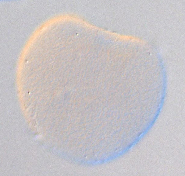
C. robusta formely Ciona int. type A, Stage 1 (One cell)

Read more…

Conversion to Aniseed format of data from Yutaka Satou's gene expression profiles of transcription factors and signalling molecule ISH screen (Development. 2004 131:4047-58).

Original annotation :
Gene name :Hox1.
Expression pattern : No signal was detected

Stained molecule KH2012:KH.L171.16 (HOXA1; HOXB1; HOXD1)
Stained region(s) No expression -

C. robusta formely Ciona int. type A, Stage 5a (early 16-cell)

Read more…

Conversion to Aniseed format of data from Yutaka Satou's gene expression profiles of transcription factors and signalling molecule ISH screen (Development. 2004 131:4047-58).

Original annotation :
Gene name :Hox1.
Expression pattern : No distinct zygotic signal was detected

Stained molecule KH2012:KH.L171.16 (HOXA1; HOXB1; HOXD1)
Stained region(s) No expression -

C. robusta formely Ciona int. type A, Stage 6b (late 32-cell)

Read more…

Conversion to Aniseed format of data from Yutaka Satou's gene expression profiles of transcription factors and signalling molecule ISH screen (Development. 2004 131:4047-58).

Original annotation :
Gene name :Hox1.
Expression pattern : No distinct zygotic signal was detected

Stained molecule KH2012:KH.L171.16 (HOXA1; HOXB1; HOXD1)
Stained region(s) No expression -

C. robusta formely Ciona int. type A, Stage 8 (64-cell)

Read more…

Conversion to Aniseed format of data from Yutaka Satou's gene expression profiles of transcription factors and signalling molecule ISH screen (Development. 2004 131:4047-58).

Original annotation :
Gene name :Hox1.
Expression pattern : No distinct zygotic signal was detected

Stained molecule KH2012:KH.L171.16 (HOXA1; HOXB1; HOXD1)
Stained region(s) No expression -

C. robusta formely Ciona int. type A, Stage 10 (112-cell)

Read more…

Conversion to Aniseed format of data from Yutaka Satou's gene expression profiles of transcription factors and signalling molecule ISH screen (Development. 2004 131:4047-58).

Original annotation :
Gene name :Hox1.
Expression pattern : No distinct zygotic signal was detected

Stained molecule KH2012:KH.L171.16 (HOXA1; HOXB1; HOXD1)
Stained region(s) No expression -

C. robusta formely Ciona int. type A, Stage 13 (late gastrula)

Read more…

Vegetal view of a late gastrula stage embryo probed for CiHox-1 expression and treated with DMSO.

Original annotation:
CiHox1 mRNA was not detected at gastrula stage.

Stained molecule KH2012:KH.L171.16 (HOXA1; HOXB1; HOXD1)
Stained region(s) No expression -

C. robusta formely Ciona int. type A, Stage 13 (late gastrula)

Read more…

Vegetal view of a late gastrula stage embryo treated with retinoic acid (RA) and probed for CiHox-1 expression. Control picture shows a late gastrula stage embryo treated with DMSO.

Original annotation:
RA strongly enhanced CiHox-1 expression. In RA-treated, the anterior ectodermal cells and anterior endodermal cells were stained (en, endoderm; ep, epidermis).

Stained molecule KH2012:KH.L171.16 (HOXA1; HOXB1; HOXD1)
Stained region(s) head endoderm - head epidermis -

C. robusta formely Ciona int. type A, Stage 14 (early neurula)

Read more…

Lateral view of an early neurula stage, DMSO-terated embryo probed for the expression of Ci Hox-1 mRNA. The dorsal side is up. nc, nerve cord.

During neurulation, the nerve cord cells in the neck region start expressing CiHox-1 mRNA.

Aniseed annotation:
Precursors of the nerve cord at the neurula stage are A10.64, A10.63, b9.37, A10.58, A10.30, A10.57, A10.29, A10.25 cell pairs.

Stained molecule KH2012:KH.L171.16 (HOXA1; HOXB1; HOXD1)
Stained region(s) A10.25 cell pair - A10.29 cell pair - A10.30 cell pair - A10.57 cell pair - A10.58 cell pair - A10.63 cell pair - A10.64 cell pair - b9.37 cell pair -

C. robusta formely Ciona int. type A, Stage 14 (early neurula)

Read more…

Expression of Ci Hox-1 mRNA in a lateral view of an early neurula stage embryo (picture 1: DMSO-treated, picture 2: RA-treated (retinoic acid)). The dorsal side is up. ep, epidermis ; en, endoderm.

Stained molecule KH2012:KH.L171.16 (HOXA1; HOXB1; HOXD1)
Stained region(s) central head endoderm (B7.1 line) - ventral head epidermis -

C. robusta formely Ciona int. type A, Stage 15 (mid neurula)

Read more…

Conversion to Aniseed format of data from Yutaka Satou's gene expression profiles of transcription factors and signalling molecule ISH screen (Development. 2004 131:4047-58).

Original annotation :
Gene name :Hox1.
Expression pattern : No distinct zygotic signal was detected

Stained molecule KH2012:KH.L171.16 (HOXA1; HOXB1; HOXD1)
Stained region(s) No expression -

C. robusta formely Ciona int. type A, Stage 17 (initial tailbud I)

Read more…

Early tailbud wild type embryos probed for the regulatory region upstream of Ci-Hox1 from -2488bp to +502bp. In this construct a part of the intron 2 of the gene was added, but there is also a deletion of 821bp (construct named Ci-Hox(deltaBsSp)/lacZ in the paper). This deletion caused the loss of the RARE regulatory motif.
Insets show similar expression patterns observed in multiple specimens.

The reporter expression was expressed in the neural tube (nt), but expression in epidermis was absent.

Stained molecule KH2012:KH.L171.16 (HOXA1; HOXB1; HOXD1)
Stained region(s) neck - tail nerve cord - visceral ganglion -

C. robusta formely Ciona int. type A, Stage 17 (initial tailbud I)

Read more…

Lateral view of an initial tailbud wild type embryo probed for reporter expression of the regulatory region upstream of Ci-Hox1 from -2488bp to +502bp. In this construct a part of the intron 2 of the gene was added (construct named Ci-Hox1(intron 2)/lacZ in the paper).
Insets show similar expression patterns observed in multiple specimens.

Regulatory region of Ci-Hox1 with added intron2 was expressed in the epidermis (ep) and neural tube (nt). Epidermal expression was restricted to the posterior half of the trunk and the anterior third of the tail in tailbud stage embryos.

Stained molecule KH2012:KH.L171.16 (HOXA1; HOXB1; HOXD1)
Stained region(s) anterior tail epidermis - dorso lateral head epidermis (a7.15 line) - neck - posterior dorsal midline head epidermis (b8.20 line) - posterior ventral head epidermis (a7.16 line) - tail nerve cord - visceral ganglion -

C. robusta formely Ciona int. type A, Stage 19 (early tailbud I)

Read more…

Dorsal views of early tailbud stage embryo expressing Hox1. The upper panel shows WISH signals, the middle DAPI signals and the lower the merged images.
Arrowheads indicate nuclei of the cells that express Hox1. Open arrows indicate the constrictions in the row of nuclei of the lateral cells. Red dots indicate nuclei of the landmark cells.

Original annotation:
The first and second domains contain four cells and at least two cells on each side, respectively. Between the two domains, the E1 and E2 cells are located.
NB: All genes described in figure 4 are expressed exclusively in the lateral cell rows s except for CiHox1, which is expressed in the dorsal and ventral cells as well.

Stained molecule KH2012:KH.L171.16 (HOXA1; HOXB1; HOXD1)
Stained region(s) neck - tail nerve cord - visceral ganglion -

C. robusta formely Ciona int. type A, Stage 19 (early tailbud I)

Read more…

Conversion to Aniseed format of data from Yutaka Satou's gene expression profiles of transcription factors and signalling molecule ISH screen (Development. 2004 131:4047-58).

Original annotation :
Gene name :Hox1.
Expression pattern : nervous system, nerve cord, epidermis.
Comment : Neural tissues and epidermal cells in the restricted region around the base of the trunk.

Stained molecule KH2012:KH.L171.16 (HOXA1; HOXB1; HOXD1)
Stained region(s) central nervous system - epidermis -

C. robusta formely Ciona int. type A, Stage 19 (early tailbud I)

Read more…

Dorsal and lateral views of an early tailbud stage, DMSO-treated embryo, probed for the expression of Ci Hox-1 mRNA. The dorsal side is up. nc, nerve cord; ep, epidermis; no , notochord.

Stained molecule KH2012:KH.L171.16 (HOXA1; HOXB1; HOXD1)
Stained region(s) posterior ventral head epidermis (a7.16 line) - tail nerve cord - ventral lateral anterior tail epidermis - visceral ganglion -

C. robusta formely Ciona int. type A, Stage 19 (early tailbud I)

Read more…

Expression of Ci Hox-1 mRNA in dorsal and lateral views of an early tailbud stage embryo (picture 1: DMSO-treated, picture 2: RA-treated (retinoic acid)) nc, nerve cord; ep, epidermis; en, endoderm; no, notochord; br, brain.

Original annotation:
In RA-treated embryo, CiHox-1 expression was observed in the entire epidermis and nerve cord cells.

Aniseed annotation:
According to the picture, it seems that the staining is also observed in the head endorderm.

Stained molecule KH2012:KH.L171.16 (HOXA1; HOXB1; HOXD1)
Stained region(s) central anterior head endoderm (A7.2 line) - central nervous system - epidermis -

C. robusta formely Ciona int. type A, Stage 19 (early tailbud I)

Read more…

Lateral and dorsal views of an early tailbud wild type embryo probed for Ci-Hox1 expression. Signal was detected in the epidermis and in the central nervous system around the junction of the trunk and the tail without clear anterior and posterior expression boundaries.

Stained molecule KH2012:KH.L171.16 (HOXA1; HOXB1; HOXD1)
Stained region(s) posterior sensory vesicle - tail nerve cord - ventral lateral anterior tail epidermis - ventral midline posterior tail epidermis - visceral ganglion -

C. robusta formely Ciona int. type A, Stage 19 (early tailbud I)

Read more…

Two-color-fluorescence WISH was performed at the early tailbud stage. Hox1 expression is shown in red, vAChTP in green.

Original annotation:
Ci-Hox1 is expressed in visceral ganglion and in part of tail nerve cord. The anterior expression domain of vAChTP is located in the gap between the two expression domains of Ci-Hox1, and the posterior domain of Ci-vAChTP is located postero-lateraly in relation to the second domain of Ci-Hox1.

Stained molecule KH2012:KH.L171.16 (HOXA1; HOXB1; HOXD1)
Stained region(s) neck - tail nerve cord - visceral ganglion -

C. robusta formely Ciona int. type A, Stage 19 (early tailbud I)

Read more…

Two-color-fluorescence WISH was performed at the early tailbud stage. Hox1 expression is shown in green, Otx in red.

Original annotation:
Ci-Hox1 is expressed in visceral ganglion and part of tail nerve cord. The first domain of Ci-Hox1 is also just behind the Ci-Otx domain without any gap.

Stained molecule KH2012:KH.L171.16 (HOXA1; HOXB1; HOXD1)
Stained region(s) neck - posterior ventral head epidermis (a7.16 line) - tail nerve cord - visceral ganglion -

C. robusta formely Ciona int. type A, Stage 19 (early tailbud I)

Read more…

Two-color fluorescence WISH was performed at the early tailbud stage. Hox1 expression is shown in green, Pax2/5/8a in red.

Original annotation:
Ci-Hox1 is expressed in visceral ganglion and in part of tail nerve cord. The anterior boundaries of the Ci-Pax2/5/8a domain and the first Ci-Hox1 domain coincide, with the former being included by the latter.

Stained molecule KH2012:KH.L171.16 (HOXA1; HOXB1; HOXD1)
Stained region(s) neck - posterior ventral head epidermis (a7.16 line) - tail nerve cord - visceral ganglion -

C. robusta formely Ciona int. type A, Stage 19 (early tailbud I)

Read more…

Two-color-fluorescence WISH was performed at the early tailbud stage. Hox1 expression is shown in red, En in green.

Original annotation:
Ci-Hox1 is expressed in visceral ganglion and in part of tail nerve cord. The second domain of Ci-En overlaps with the posterior part of the first domain of Hox1, while the first domain of Ci-En is just ahead of the first domain of Ci-Hox1.

Stained molecule KH2012:KH.L171.16 (HOXA1; HOXB1; HOXD1)
Stained region(s) neck - posterior ventral head epidermis (a7.16 line) - tail nerve cord - visceral ganglion -

C. robusta formely Ciona int. type A, Stage 19 (early tailbud I)

Read more…

Two-color-fluorescence WISH was performed at the early tailbud stage. Hox1 expression is shown in red, SoxB1 in green.

Original annotation:
Ci-Hox1 is expressed in visceral ganglion and in part of tail nerve cord. The second domain of Ci-SoxB1 overlaps with the anterior part of the second domain of Ci-Hox1.

Stained molecule KH2012:KH.L171.16 (HOXA1; HOXB1; HOXD1)
Stained region(s) neck - posterior ventral head epidermis (a7.16 line) - tail nerve cord - visceral ganglion -

C. robusta formely Ciona int. type A, Stage 19 (early tailbud I)

Read more…

Two-color-fluorescence WISH was performed at the early tailbud stage. Hox1 expression is shown in red, Lhx3 in green.

Original annotation:
Ci-Hox1 is expressed in visceral ganglion and in part of tail nerve cord. The anterior expression domain of Ci-Lhx3 is located in the gap between the two expression domains of Ci-Hox1, and the posterior domain of Ci-Lhx3 is located postero-lateraly in relation to the second domain of Ci-Hox1.

Stained molecule KH2012:KH.L171.16 (HOXA1; HOXB1; HOXD1)
Stained region(s) neck - tail nerve cord - visceral ganglion -

C. robusta formely Ciona int. type A, Stage 19 (early tailbud I)

Read more…

Two-color-fluorescence WISH was performed at the early tailbud stage. Hox1 expression is shown in green, Hox5 in red.

Original annotation: Ci-Hox1 is expressed in visceral ganglion and in part of tail nerve cord. the anterior expression boundary of Ci-Hox5 is posterior to that of the second domain of Ci-Hox1.

Stained molecule KH2012:KH.L171.16 (HOXA1; HOXB1; HOXD1)
Stained region(s) neck - posterior ventral head epidermis (a7.16 line) - tail nerve cord - visceral ganglion -

C. robusta formely Ciona int. type A, Stage 19 (early tailbud I)

Read more…

Lateral view of an early tailbud stage embryo showing the expression pattern of citb14p24 clone for which expression was activated by retinoic acid. The embryos were treated with DMSO. Arrows indicate the staining in the anterior tail epidermis.

Original annotation:
In control embryos, 01890r1 was expressed in the epidermis and nerve cord of the anterior tail region.

Stained molecule KH2012:KH.L171.16 (HOXA1; HOXB1; HOXD1)
Stained region(s) anterior tail epidermis - tail nerve cord -

C. robusta formely Ciona int. type A, Stage 19 (early tailbud I)

Read more…

Vegetal view of a late gastrula stage embryo showing the expression pattern of a selected clone (citb14p24) for which expression was activated by retinoic acid. The embryos were treated with retinoic acid (RA).

Original annotation:
01890r1 was activated by RA in the entire ectodermal tissues, including the central nervous system.

Stained molecule KH2012:KH.L171.16 (HOXA1; HOXB1; HOXD1)
Stained region(s) central nervous system - epidermis -

C. robusta formely Ciona int. type A, Stage 19 (early tailbud I)

Read more…

Dorsal view of an early tailbud wild type embryo probed for Ci-Hox1 expression. Red and yellow arrowheads indicate boundaries between the neck and the visceral ganglion and between the visceral ganglion and the caudal nerve cord, respectively.

Original Annotation :
Ci-Hox1 was expressed in A11.61, A11.62 cell pairs (neck), A11.119, A11.120 cell pairs (visceral ganglion) and in A11.115, A11.116 cell pairs (lateral nerve cord).

Stained molecule KH2012:KH.L171.16 (HOXA1; HOXB1; HOXD1)
Stained region(s) neck - tail nerve cord - visceral ganglion -

C. robusta formely Ciona int. type A, Stage 19 (early tailbud I)

Read more…

Dorsal view of an early tailbud wild type embryo probed for Ci-Hox1 IN Raldh2 knockdown embryos.
Red and yellow arrowheads indicate boundaries between the neck and the visceral ganglion and between the visceral ganglion and the caudal nerve cord, respectively.

Original Annotation :
Expression was lost.

Stained molecule KH2012:KH.L171.16 (HOXA1; HOXB1; HOXD1)
Stained region(s) No expression -

C. robusta formely Ciona int. type A, Stage 19 (early tailbud I)

Read more…

Dorsal view of an early tailbud wild type embryo probed for Ci-Hox1 expression. The insert shows in red cells from the posterior sensory vesicle (PSV) and light-blue cells from the middle part of the visceral ganglion (VG). Broken white lines indicate the boundaries of the PSV/neck and the neck/VG.

Original Annotation :
Ci-Hox1 expression was detected in A11.62 and A11.61 cell pairs (neck) and in A11.120 and A11.119 cell pairs (anterior visceral ganglion).

Stained molecule KH2012:KH.L171.16 (HOXA1; HOXB1; HOXD1)
Stained region(s) neck - visceral ganglion -

C. robusta formely Ciona int. type A, Stage 19 (early tailbud I)

Read more…

Dorsal view of an early tailbud embryo probed for Ci-Hox1 expression, following the injection of Fgf8/17/18 MO.
The insert shows in red cells from the posterior sensory vesicle (PSV) and light-blue cells from the middle part of the visceral ganglion (VG). Broken white lines indicate the boundaries of the PSV/neck and the neck/VG.

Original Annotation :
Ci-Hox1 expression in the neck was lost.

Stained molecule KH2012:KH.L171.16 (HOXA1; HOXB1; HOXD1)
Stained region(s) visceral ganglion -

C. robusta formely Ciona int. type A, Stage 19 (early tailbud I)

Read more…

Lateral view of an early tailbud wild type embryo probed for reporter expression of the regulatory region upstream of Ci-Hox1 from -2488bp to +502bp.
In this construct a part of the intron 2 of the gene was added, and the RARE regulatory motif in the sequence was mutated (construct named Ci-Hox1(intron 2)mut/lacZ in the paper).
Insets show similar expression patterns observed in multiple specimens.

Regulatory region of Ci-Hox1 with the intron2 added was expressed in the neural tube (nt). The expression in epidermis was lost, due to the mutation of the RARE site.

Stained molecule KH2012:KH.L171.16 (HOXA1; HOXB1; HOXD1)
Stained region(s) neck - tail nerve cord - visceral ganglion -

C. robusta formely Ciona int. type A, Stage 19 (early tailbud I)

Read more…

Lateral view of an early tailbud wild type embryo probed for reporter expression of the regulatory region upstream of Ci-Hox1 from -2488bp to +502bp. In this construct a part of the intron 2 of the gene was added (construct named Ci-Hox1(intron 2)/lacZ in the paper).
Insets show similar expression patterns observed in multiple specimens.

Regulatory region of Ci-Hox1 with added intron2 was expressed in the epidermis (ep) and neural tube (nt). Epidermal expression was restricted to the posterior half of the trunk and the anterior third of the tail in tailbud stage embryos.

Stained molecule KH2012:KH.L171.16 (HOXA1; HOXB1; HOXD1)
Stained region(s) anterior tail epidermis - dorso lateral head epidermis (a7.15 line) - neck - posterior dorsal midline head epidermis (b8.20 line) - posterior ventral head epidermis (a7.16 line) - tail nerve cord - visceral ganglion -

C. robusta formely Ciona int. type A, Stage 19 (early tailbud I)

Read more…

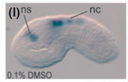
Original annotation: expression of the transgene in nerve cord. Expression restricted to the visceral ganglion and anterior nerve cord region (similar to endogenous Hox1 mRNA). ns, non specific expression ; nc, nerve cord.

Stained molecule KH2012:KH.L171.16 (HOXA1; HOXB1; HOXD1)
Stained region(s) tail nerve cord - visceral ganglion -

C. robusta formely Ciona int. type A, Stage 19 (early tailbud I)

Read more…

lateral view of embryos.
Original annotation: similar pattern than the full length intron 2 construct and endogenous Hox1 mRNA. Expression restricted to the visceral ganglion and anterior nerve cord region.
nc, nerve cord ; ns, non specific expression

Stained molecule KH2012:KH.L171.16 (HOXA1; HOXB1; HOXD1)
Stained region(s) tail nerve cord - visceral ganglion -

C. robusta formely Ciona int. type A, Stage 19 (early tailbud I)

Read more…

lateral view of embryos. Original annotation: similar pattern than the full length intron 2 construct and endogenous Hox1 mRNA. Expression restricted to the visceral ganglion and anterior nerve cord region.
nc, nerve cord ; ns, non specific expression

Stained molecule KH2012:KH.L171.16 (HOXA1; HOXB1; HOXD1)
Stained region(s) tail nerve cord - visceral ganglion -

C. robusta formely Ciona int. type A, Stage 19 (early tailbud I)

Read more…

Lateral view of embryo.
Original annotation: The transgene containing 1828 bp of the intronic sequence (Ci-Hox1(i2D4)/lacZ) was not expressed in the nerve cord.
ns, non specific expression (mesenchyme).

Stained molecule KH2012:KH.L171.16 (HOXA1; HOXB1; HOXD1)
Stained region(s)

C. robusta formely Ciona int. type A, Stage 19 (early tailbud I)

Read more…

Lateral view of an early tailbud stage embryo stained with X-gal electroporated with the RA reporter construct pCi-Hox(intron)::LacZ.
Original annotation: Expression was observed in the anterior tail epidermis (bracket) and in some muscle cells (black arrowheads).

Stained molecule KH2012:KH.L171.16 (HOXA1; HOXB1; HOXD1)
Stained region(s) anterior tail epidermis - mesenchyme (B8.5 & B7.7 lines) - neck - posterior dorsal midline head epidermis (b8.20 line) - posterior ventral head epidermis (a7.16 line) - tail nerve cord - visceral ganglion -

C. robusta formely Ciona int. type A, Stage 19 (early tailbud I)

Read more…

Lateral view of an early tailbud stage embryo stained with X-gal electroporated with the RA reporter construct pCi-Hox(intron)::LacZ after retinoic acide treatment (1.5μM) at the late gastrula stage.
Original annotation: Ectopic expression was observed throughout the embryo.

Stained molecule KH2012:KH.L171.16 (HOXA1; HOXB1; HOXD1)
Stained region(s) whole embryo -

C. robusta formely Ciona int. type A, Stage 20 (early tailbud II)

Read more…

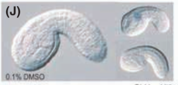
Lateral view of embryo.
Original annotation: Weak expression in the nerve cord. nc, nerve cord; ns, non-specific expression.

Stained molecule KH2012:KH.L171.16 (HOXA1; HOXB1; HOXD1)
Stained region(s) tail nerve cord -

C. robusta formely Ciona int. type A, Stage 20 (early tailbud II)

Read more…

Lateral view of embryo.
Original annotation: Weak expression in the nerve cord. nc, nerve cord.

Stained molecule KH2012:KH.L171.16 (HOXA1; HOXB1; HOXD1)
Stained region(s) tail nerve cord -

C. robusta formely Ciona int. type A, Stage 20 (early tailbud II)

Read more…

Lateral view of embryo.
Original annotation: No expression in the nerve cord. ns, non-specific expression (mesenchyme).

Stained molecule KH2012:KH.L171.16 (HOXA1; HOXB1; HOXD1)
Stained region(s)

C. robusta formely Ciona int. type A, Stage 20 (early tailbud II)

Read more…

Lateral view of embryo.
Original annotation: No expression in the nerve cord or the whole embryo.
Aniseed annotation: maybe a weak non specific expression in embryo on the upper right panel.

Stained molecule KH2012:KH.L171.16 (HOXA1; HOXB1; HOXD1)
Stained region(s) No expression -

C. robusta formely Ciona int. type A, Stage 20 (early tailbud II)

Read more…

Lateral view of embryo.
Original annotation: Expression in the nerve cord. nc, nerve cord.

Stained molecule KH2012:KH.L171.16 (HOXA1; HOXB1; HOXD1)
Stained region(s) tail nerve cord -

C. robusta formely Ciona int. type A, Stage 21 (mid tailbud I)

Read more…

Lateral view of embryo. Experiment on tailbud embryos developed from eggs injected with lacZ-specific control MO.
Original annotation: Strong expression in the nerve cord and epidermis. nc, nerve cord; ep, epidermis.

Stained molecule KH2012:KH.L171.16 (HOXA1; HOXB1; HOXD1)
Stained region(s) anterior tail epidermis - tail nerve cord -

C. robusta formely Ciona int. type A, Stage 21 (mid tailbud I)

Read more…

Dorsal views of mid tailbud stage embryo expressing Hox1. The upper panel shows WISH signals, the middle DAPI signals and the lower the merged images.
Arrowheads indicate nuclei of the cells that express Hox1. Open arrows indicate the constrictions in the row of nuclei of the lateral cells. Red dots indicate nuclei of the landmark cells.

Original annotation:
The fourth cell from the anterior in the first domain of Ci-Hox1 corresponds to the M1 cell, and two cells between the two domains are the M2 and M3 cells.
NB: All genes described in figure 4 are expressed exclusively in the lateral cell rows except for CiHox1, which is expressed in the dorsal and ventral cells as well.

Stained molecule KH2012:KH.L171.16 (HOXA1; HOXB1; HOXD1)
Stained region(s) neck - tail nerve cord - visceral ganglion -

C. robusta formely Ciona int. type A, Stage 21 (mid tailbud I)

Read more…

Conversion to Aniseed format of data from Yutaka Satou's gene expression profiles of transcription factors and signalling molecule ISH screen (Development. 2004 131:4047-58).

Original annotation :
Gene name :Hox1.
Expression pattern : nervous system, nerve cord, epidermis.
Comment : Neural tissues and epidermal cells in the restricted region around the base of the trunk.

Stained molecule KH2012:KH.L171.16 (HOXA1; HOXB1; HOXD1)
Stained region(s) central nervous system - posterior ventral head epidermis (a7.16 line) - ventral lateral anterior tail epidermis - ventral midline anterior tail epidermis -

C. robusta formely Ciona int. type A, Stage 21 (mid tailbud I)

Read more…

Lateral view of a mid tailbud stage wild type embryo probed for Ci-Hox1 expression.

Stained molecule KH2012:KH.L171.16 (HOXA1; HOXB1; HOXD1)
Stained region(s) posterior sensory vesicle - posterior ventral head epidermis (a7.16 line) - tail nerve cord - ventral lateral anterior tail epidermis - ventral midline anterior tail epidermis - visceral ganglion -

C. robusta formely Ciona int. type A, Stage 21 (mid tailbud I)

Read more…

Two-color-fluorescence WISH was performed at the mid tailbud stage. Hox1 expression is shown in green, Otx in red.

Original annotation:
Ci-Hox1 is expressed in visceral ganglion and part of tail nerve cord. There is a gap between the Ci-Otx domain and the first domain of Ci-Hox1.

Stained molecule KH2012:KH.L171.16 (HOXA1; HOXB1; HOXD1)
Stained region(s) neck - tail nerve cord - ventral midline anterior tail epidermis - visceral ganglion -

C. robusta formely Ciona int. type A, Stage 21 (mid tailbud I)

Read more…

Two-color-fluorescence WISH was performed at the mid tailbud stage. Hox1 expression is shown in green, Pax2/5/8a in red.

Original annotation:
Ci-Hox1 is expressed in visceral ganglion and part of tail nerve cord. The anterior part of the first domain of Ci-Hox1 overlaps with the posterior part of the Ci-Pax2/5/8a domain.

Stained molecule KH2012:KH.L171.16 (HOXA1; HOXB1; HOXD1)
Stained region(s) neck - tail nerve cord - ventral midline anterior tail epidermis - visceral ganglion -

C. robusta formely Ciona int. type A, Stage 21 (mid tailbud I)

Read more…

Two-color-fluorescence WISH was performed at the mid tailbud stage. Hox1 expression is shown in red, En in red.

Original annotation:
Ci-Hox1 is expressed in visceral ganglion and in part of tail nerve cord. The second and third domains of Ci-En overlap with the anterior and posterior parts of the first domain of Ci-Hox1.

Stained molecule KH2012:KH.L171.16 (HOXA1; HOXB1; HOXD1)
Stained region(s) neck - tail nerve cord - visceral ganglion -

C. robusta formely Ciona int. type A, Stage 21 (mid tailbud I)

Read more…

Two-color-fluorescence WISH was performed at the mid tailbud stage. Hox1 expression is shown in red, Fgf8/17/18 in green.

Original annotation:
Ci-Hox1 is expressed in visceral ganglion and partg of tail nerve cord. Expression of Ci-Fgf8/17/18 was detected in a small region of the CNS, which overlaps with posterior part of the first domain of Ci-Hox1.

Stained molecule KH2012:KH.L171.16 (HOXA1; HOXB1; HOXD1)
Stained region(s) neck - tail nerve cord - ventral lateral anterior tail epidermis - visceral ganglion -

C. robusta formely Ciona int. type A, Stage 21 (mid tailbud I)

Read more…

Two-color-fluorescence WISH was performed at the mid tailbud stage. Hox1 expression is shown in red, SoxB1 in green.

Original annotation:
Ci-Hox1 is expressed in visceral ganglion and in part of tail nerve cord. The expression domain of Ci-SoxB1 in the anterior nerve cord is included by the second domain of Ci-Hox1, which expands more posteriorly without distinct boundary.

Stained molecule KH2012:KH.L171.16 (HOXA1; HOXB1; HOXD1)
Stained region(s) neck - posterior ventral head epidermis (a7.16 line) - tail nerve cord - ventral midline anterior tail epidermis - visceral ganglion -

C. robusta formely Ciona int. type A, Stage 21 (mid tailbud I)

Read more…

Two-color-fluorescence WISH was performed at the mid tailbud stage. Hox1 expression is shown in red, vACHTP in green.

Original annotation:
Ci-Hox1 is expressed in the visceral ganglion and in part of tail nerve cord. Ci-vACHTP is expressed in the gap between the two Ci-Hox1 domains as at the early stage and in the domain just ventrolateral to the second domain of Ci-Hox1.

Stained molecule KH2012:KH.L171.16 (HOXA1; HOXB1; HOXD1)
Stained region(s) neck - tail nerve cord - ventral lateral anterior tail epidermis - ventral midline anterior tail epidermis - visceral ganglion -

C. robusta formely Ciona int. type A, Stage 21 (mid tailbud I)

Read more…

Two-color-fluorescence WISH was performed at the mid tailbud stage. Hox1 expression is shown in red, Lhx3 in green.

Original annotation:
Ci-Hox1 is expressed in the visceral ganglion and in part of tail nerve cord. The anterior domain of Ci-Lhx3 extends anteriorly up to the posterior part of the first Ci-Hox1 domain.

Stained molecule KH2012:KH.L171.16 (HOXA1; HOXB1; HOXD1)
Stained region(s) neck - tail nerve cord - ventral midline anterior tail epidermis - visceral ganglion -

C. robusta formely Ciona int. type A, Stage 21 (mid tailbud I)

Read more…

Two-color-fluorescence WISH was performed at the mid tailbud stage. Hox1 expression is shown in red, Hox5 in green. Insert indicates higher magnification of the second domain of Ci-Hox1 and the anterior part of Ci-Hox5 domain. Images of DAPI staining is additionally merged.

Original annotation:
Ci-Hox1 is expressed in visceral ganglion and in part of tail nerve cord. In the tail nerve cord, the anterior expression boundary of Ci-Hox5 is behind that of the second domain of Ci-Hox1 by two cells.

Stained molecule KH2012:KH.L171.16 (HOXA1; HOXB1; HOXD1)
Stained region(s) neck - tail nerve cord - visceral ganglion -

C. robusta formely Ciona int. type A, Stage 21 (mid tailbud I)

Read more…

Dorsal view of a mid tailbud wild type embryo probed for Hox1 expression.
Red and yellow arrowheads indicate boundaries between the neck and the visceral ganglion, and between the visceral ganglion and the caudal nerve cord, respectively.

Original Annotation :
Hox1 was expressed in the visceral ganglion (more precisely in A12.238, A12.239 A12.240, A13.473, A13.474 cell pairs), in the neck and in the lateral nerve cord.

Stained molecule KH2012:KH.L171.16 (HOXA1; HOXB1; HOXD1)
Stained region(s) lateral TNC ependymal precursors - neck - visceral ganglion -

C. robusta formely Ciona int. type A, Stage 21 (mid tailbud I)

Read more…

Dorsal view of a mid tailbud embryo probed for Hox1 expression following the injection of FGF8/17/18 MO.
Red and yellow arrowheads indicate boundaries between the neck and the visceral ganglion, and between the visceral ganglion and the caudal nerve cord, respectively.

Original Annotation :
Hox1 expression was lost in the neck (white arrow).

Stained molecule KH2012:KH.L171.16 (HOXA1; HOXB1; HOXD1)
Stained region(s) lateral TNC ependymal precursors - visceral ganglion -

C. robusta formely Ciona int. type A, Stage 21 (mid tailbud I)

Read more…

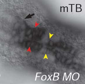
Dorsal view of a mid tailbud embryo probed for Hox1 expression following the injection of FoxB MO.
Red and yellow arrowheads indicate boundaries between the neck and the visceral ganglion, and between the visceral ganglion and the caudal nerve cord, respectively.

Original Annotation :
Ectopic expression was observed (black arrow).

Aniseed Annotation :
According to the picture, expression seems to be in the posterior sensory vesicle

Stained molecule KH2012:KH.L171.16 (HOXA1; HOXB1; HOXD1)
Stained region(s) lateral TNC ependymal precursors - neck - posterior sensory vesicle - visceral ganglion -

C. robusta formely Ciona int. type A, Stage 21 (mid tailbud I)

Read more…

Dorsal view of a mid tailbud embryo probed for Hox1 expression following the injection of Neurogenin MO.
Red and yellow arrowheads indicate boundaries between the neck and the visceral ganglion, and between the visceral ganglion and the caudal nerve cord, respectively.

Original Annotation :
Ectopic expression was observed (black arrow).

Aniseed Annotation :
According to the picture, ectopic expression is localized in the nerve cord.

Stained molecule KH2012:KH.L171.16 (HOXA1; HOXB1; HOXD1)
Stained region(s) neck - tail nerve cord - visceral ganglion -

C. robusta formely Ciona int. type A, Stage 21 (mid tailbud I)

Read more…

Dorsal view of a mid tailbud embryo probed for Hox1 expression following the injection of Otx MO.
Red and yellow arrowheads indicate boundaries between the neck and the visceral ganglion, and between the visceral ganglion and the caudal nerve cord, respectively.

Original Annotation :
Ectopic expression was observed (black arrow).

Aniseed Annotation :
According to the picture, ectopic expression seems to be localized in the neck.

Stained molecule KH2012:KH.L171.16 (HOXA1; HOXB1; HOXD1)
Stained region(s) lateral TNC ependymal precursors - neck - visceral ganglion -

C. robusta formely Ciona int. type A, Stage 21 (mid tailbud I)

Read more…

Mid tailbud wild type embryos probed for the reporter expression of the regulatory region upstream of Ci-Hox1 from -2488bp to +502bp. In this construct a part of the intron 2 of the gene was added (construct named Ci-Hox1(intron 2)/lacZ in the paper).
These embryos were treated with EtOH as control.
Insets show similar expression patterns observed in multiple specimens.

Embryos showed expression in neural tube (neck, visceral ganglion, part of anterior tail nerve cord) and in the epidermis (posterior trunk epidermis, and anterior tail epidermis).

Stained molecule KH2012:KH.L171.16 (HOXA1; HOXB1; HOXD1)
Stained region(s) anterior tail epidermis - dorso lateral head epidermis (a7.15 line) - neck - posterior dorsal midline head epidermis (b8.20 line) - posterior ventral head epidermis (a7.16 line) - tail nerve cord - visceral ganglion -

C. robusta formely Ciona int. type A, Stage 21 (mid tailbud I)

Read more…

Regulatory region upstream of Ci-Hox1 from -2488bp to +502bp. In this construct a part of the intron 2 of the gene was added (construct named Ci-Hox1(intron 2)/lacZ in the paper).
Insets show similar expression patterns observed in multiple specimens.

Regulatory region of Ci-Hox1 with the intron2 added was expressed in the epidermis (ep) and neural tube (nt). Epidermal expression was restricted to the posterior half of the trunk and the anterior third of the tail in tailbud stage embryos

Stained molecule KH2012:KH.L171.16 (HOXA1; HOXB1; HOXD1)
Stained region(s) anterior tail epidermis - dorso lateral head epidermis (a7.15 line) - neck - posterior dorsal midline head epidermis (b8.20 line) - posterior ventral head epidermis (a7.16 line) - tail nerve cord - visceral ganglion -

C. robusta formely Ciona int. type A, Stage 21 (mid tailbud I)

Read more…

Regulatory region upstream of Ci-Hox1 from -2488bp to +502bp. In this construct a part of the intron 2 of the gene was added (construct named Ci-Hox1(intron 2)/lacZ in the paper).
Insets show similar expression patterns observed in multiple specimens.

Original Annotation : A fluorescent staining using a β-galactosidase-specific antibody revealed that the expression in the neural tube was restricted to the neck, visceral ganglion and anterior nerve cord regions.

Stained molecule KH2012:KH.L171.16 (HOXA1; HOXB1; HOXD1)
Stained region(s) dorsal midline anterior tail epidermis - neck - tail nerve cord - visceral ganglion -

C. robusta formely Ciona int. type A, Stage 21 (mid tailbud I)

Read more…

Regulatory region upstream of Ci-Hox1 from -2488bp to +502bp. In this construct a part of the intron 2 of the gene was added (construct named Ci-Hox1(intron 2)/lacZ in the paper). These embryos were treated with DMSO as control.
Insets show similar expression patterns observed in multiple specimens.

Regulatory region of Ci-Hox1 with the intron2 added was expressed in the epidermis (ep) and neural tube, more precisely in the neck, visceral ganglion and anterior nerve cord. Epidermal expression was restricted to the posterior half of the trunk and the anterior third of the tail in tailbud stage embryos

Stained molecule KH2012:KH.L171.16 (HOXA1; HOXB1; HOXD1)
Stained region(s) anterior tail epidermis - dorso lateral head epidermis (a7.15 line) - neck - posterior ventral head epidermis (a7.16 line) - tail nerve cord - visceral ganglion -

C. robusta formely Ciona int. type A, Stage 21 (mid tailbud I)

Read more…

Regulatory region upstream of Ci-Hox1 from -2488bp to +502bp. In this construct a part of the intron 2 of the gene was added, but there is also a deletion of 821bp (construct named Ci-Hox(deltaBsSp)/lacZ in the paper). This deletion caused the loss of the RARE regulatory motif.
Insets show similar expression patterns observed in multiple specimens.

The reporter expression was expressed in the neural tube, but expression in epidermis was very weak comparing with the embryos electroporated with the construct without the 821bp deletion.

Stained molecule KH2012:KH.L171.16 (HOXA1; HOXB1; HOXD1)
Stained region(s) neck - tail nerve cord - visceral ganglion -

C. robusta formely Ciona int. type A, Stage 21 (mid tailbud I)

Read more…

Representation of the construct (named Ci-Hox1(del2)/LacZ in the paper) used to report the expression of the cis regulatory region upstream of Ci-Hox1 from -836 to 553. The exons of Ci-Hox1 are shown by gray boxes. The transcription start site is indicated by an arrow. The element 1 is indicated by a blue oval. The second element was deleted and with it the DR-2 type RARE regulatory motif.

Due to the deletion, there is a complete loss of epidermal expression (in all embryos tested). Ectopic expression appears.

Stained molecule KH2012:KH.L171.16 (HOXA1; HOXB1; HOXD1)
Stained region(s) No expression -

C. robusta formely Ciona int. type A, Stage 21 (mid tailbud I)

Read more…

Representation of the construct (named Ci-Hox1(0,9)/LacZ in the paper) used to report the expression of the cis regulatory region upstream of Ci-Hox1 from -836 to 553.
The exons of Ci-Hox1 are shown by gray boxes. The transcription start site is indicated by an arrow. Two conserved sequence elements between Ciona species are indicated by a blue oval (element 1) and an orange oval (element 2).

Reporter LacZ expression was detected in the epidermis. (in 60% of embryos probed)

Stained molecule KH2012:KH.L171.16 (HOXA1; HOXB1; HOXD1)
Stained region(s) epidermis -

C. robusta formely Ciona int. type A, Stage 21 (mid tailbud I)

Read more…

Representation of the construct (named Ci-Hox1(del1)/LacZ in the paper) used to report the expression of the cis regulatory region upstream of Ci-Hox1 from -836 to 553.
The exons of Ci-Hox1 are shown by gray boxes. The transcription start site is indicated by an arrow. The element 2 is indicated by an orange oval. The first element was deleted.

Reporter LacZ expression was detected in the epidermis (in 71% of embryos probed). The deletion has no effect on epidermal expression.

Stained molecule KH2012:KH.L171.16 (HOXA1; HOXB1; HOXD1)
Stained region(s) epidermis -

C. robusta formely Ciona int. type A, Stage 21 (mid tailbud I)

Read more…

Representation of the construct (named Ci-Hox1(0,9)mut/LacZ in the paper) used to report the expression of the cis regulatory region upstream of Ci-Hox1 from -836 to 553.
The exons of Ci-Hox1 are shown by gray boxes. The transcription start site is indicated by an arrow. The element 1 is indicated by a blue oval. The DR-2 type RARE was mutated and indicated by a red cross.

This mutation caused a loss of epidermal expression in all embryos. Ectopic expression is detected.

Stained molecule KH2012:KH.L171.16 (HOXA1; HOXB1; HOXD1)
Stained region(s) No expression -

C. robusta formely Ciona int. type A, Stage 21 (mid tailbud I)

Read more…

Mid tailbud wild type embryos probed for the reporter expression of the regulatory region upstream of Ci-Hox1 from -2488bp to +502bp. In this construct a part of the intron 2 of the gene was added (construct named Ci-Hox1(intron 2)/lacZ in the paper).
These embryos were treated with DMSO as control.
Insets show similar expression patterns observed in multiple specimens.

Embryos showed expression in neural tube (neck, visceral ganglion, part of anterior tail nerve cord) and in the epidermis (posterior trunk epidermis, and anterior tail epidermis).

Stained molecule KH2012:KH.L171.16 (HOXA1; HOXB1; HOXD1)
Stained region(s) anterior tail epidermis - dorso lateral head epidermis (a7.15 line) - neck - posterior dorsal midline head epidermis (b8.20 line) - posterior ventral head epidermis (a7.16 line) - tail nerve cord - visceral ganglion -

C. robusta formely Ciona int. type A, Stage 22 (mid tailbud II)

Read more…

Lateral view with anterior to the left of a mid-tailbud stage embryo probed for Hox1 expression.
Original annotation: Expression was observed in the anterior third tail epidermal cells.

Stained molecule KH2012:KH.L171.16 (HOXA1; HOXB1; HOXD1)
Stained region(s) posterior dorsal midline head epidermis (b8.20 line) - posterior ventral head epidermis (a7.16 line) - tail nerve cord - ventral lateral anterior tail epidermis - ventral midline anterior tail epidermis - visceral ganglion -

C. robusta formely Ciona int. type A, Stage 22 (mid tailbud II)

Read more…

Lateral view with anterior to the left of a mid-tailbud stage embryo stained with Hox1 after retinoic acide treatment (1.5μM) at the late gastrula stage.
Original annotation: Ectopic expression was observed throughout the tail epidermis.

Stained molecule KH2012:KH.L171.16 (HOXA1; HOXB1; HOXD1)
Stained region(s) tail epidermis - whole embryo -

C. robusta formely Ciona int. type A, Stage 22 (mid tailbud II)

Read more…

Lateral view with anterior to the left of a mid-tailbud stage embryo stained with Hox11 after DEAB treatment (150μM) at the late gastrula stage.
Original annotation: Expression was lost in the anterior third tail epidermal cells.

Stained molecule KH2012:KH.L171.16 (HOXA1; HOXB1; HOXD1)
Stained region(s) No expression -

C. robusta formely Ciona int. type A, Stage 22 (mid tailbud II)

Read more…

Lateral view with anterior to the left of a mid-tailbud stage embryo stained with Hox1 after bFGF treatment (100ng/ml) at the late gastrula stage.
Original annotation: Expression was lost in the anterior tail epidermis.

Stained molecule KH2012:KH.L171.16 (HOXA1; HOXB1; HOXD1)
Stained region(s) tail nerve cord - visceral ganglion -

C. robusta formely Ciona int. type A, Stage 22 (mid tailbud II)

Read more…

Lateral view with anterior to the left of a mid-tailbud stage embryo probed for Hox1 expression after injection of a dominant-negative form of the Ciona FGF receptor (Ci-FGFR) at the late gastrula stage.
Original annotation: Ectopic expression was observed in the tail (black arrowhead).

Stained molecule KH2012:KH.L171.16 (HOXA1; HOXB1; HOXD1)
Stained region(s) posterior dorsal midline head epidermis (b8.20 line) - posterior ventral head epidermis (a7.16 line) - tail nerve cord - ventral lateral anterior tail epidermis - ventral midline anterior tail epidermis - visceral ganglion -

C. robusta formely Ciona int. type A, Stage 22 (mid tailbud II)

Read more…

Lateral view with anterior to the left of a mid-tailbud stage embryo probed for Hox1 expression after U0126 treatment (10μM) at the late gastrula stage.
Original annotation: Expression was lost in the anterior third tail epidermal cells.

Stained molecule KH2012:KH.L171.16 (HOXA1; HOXB1; HOXD1)
Stained region(s) No expression -

C. robusta formely Ciona int. type A, Stage 23 (late tailbud I)

Read more…

Dorsal views of late tailbud stage embryo expressing Hox1. The upper panel shows WISH signals, the middle DAPI signals and the lower the merged images.
Arrowheads indicate nuclei of the cells that express Hox1. Open arrows indicate the constrictions in the row of nuclei of the lateral cells. Red dots indicate nuclei of the landmark cells.

Original annotation:
The fourth cell from the anterior in the first domain of Ci-Hox1 is the L1 cell, and two cells between the two domains are the L2 and L3 cells.
NB: All genes described in figure 4 are expressed exclusively in the lateral cell rows except for CiHox1, which is expressed in the dorsal and ventral cells as well.

Stained molecule KH2012:KH.L171.16 (HOXA1; HOXB1; HOXD1)
Stained region(s) neck - tail nerve cord - visceral ganglion -

C. robusta formely Ciona int. type A, Stage 23 (late tailbud I)

Read more…

Lateral view of a late tailbud wild type embryo probed for Ci-Hox1 expression.

Stained molecule KH2012:KH.L171.16 (HOXA1; HOXB1; HOXD1)
Stained region(s) neck - posterior sensory vesicle - posterior ventral head epidermis (a7.16 line) - tail nerve cord - ventral lateral posterior tail epidermis - ventral midline posterior tail epidermis - visceral ganglion -

C. robusta formely Ciona int. type A, Stage 23 (late tailbud I)

Read more…

Two-color-fluorescence WISH was performed at the late tailbud stage. Hox1 expression is shown in green, Otx in red.

Original annotation:
Ci-Hox1 is expressed in visceral ganglion and in part of tail nerve cord. Ci-Otx expression extends caudally along the ventral midline of the neural tube.

Stained molecule KH2012:KH.L171.16 (HOXA1; HOXB1; HOXD1)
Stained region(s) neck - posterior ventral head epidermis (a7.16 line) - tail nerve cord - ventral midline anterior tail epidermis - visceral ganglion -

C. robusta formely Ciona int. type A, Stage 23 (late tailbud I)

Read more…

Two-color-fluorescence WISH was performed at the late tailbud stage. Hox1 expression is shown in green, Pax2/5/8a in red.

Original annotation:
Ci-Hox1 is expressed in visceral ganglion and in part of tail nerve cord. Ci-Pax2/5/8a domain posteriorly overlaps with the anterior part of the first domain of Ci-Hox1.

Stained molecule KH2012:KH.L171.16 (HOXA1; HOXB1; HOXD1)
Stained region(s) neck - posterior ventral head epidermis (a7.16 line) - tail nerve cord - ventral midline anterior tail epidermis - visceral ganglion -

C. robusta formely Ciona int. type A, Stage 23 (late tailbud I)

Read more…

Two-color-fluorescence WISH was performed at the late tailbud stage. Hox1 expression is shown in red, En in green.

Original annotation:
Ci-Hox1 is expressed in visceral ganglion and part of tail nerve cord. Expression of Ci-En was detected in three domains. The posterior-most part of the first domain of Ci-Hox1 expression domain do not overlap with the third domain of Ci-En.

Stained molecule KH2012:KH.L171.16 (HOXA1; HOXB1; HOXD1)
Stained region(s) neck - posterior ventral head epidermis (a7.16 line) - tail nerve cord - ventral midline anterior tail epidermis - visceral ganglion -

C. robusta formely Ciona int. type A, Stage 23 (late tailbud I)

Read more…

Two-color-fluorescence WISH was performed at the late tailbud stage. Hox1 expression is shown in red, SoxB1 in green.

Original annotation :
Ci-Hox1 is expressed in the visceral ganglion and part of tail nerve cord. Expression of Ci-Hox1 and Ci-SoxB1 overlap in nervous system.

Stained molecule KH2012:KH.L171.16 (HOXA1; HOXB1; HOXD1)
Stained region(s) neck - tail nerve cord - ventral midline anterior tail epidermis - visceral ganglion -

C. robusta formely Ciona int. type A, Stage 23 (late tailbud I)

Read more…

Two-color-fluorescence WISH was performed at the late tailbud stage. Hox1 expression is shown in red, vAChTP in green.

Original annotation:
Ci-Hox1 is expressed in the visceral ganglion and part of tail nerve cord. At this stage, vAChTP is expressed in two distinct domains aligned along the A-P axis. While the posterior domain is located in the gap between the two domains of Ci-Hox1 continuously, the anterior domain lies the middle of the first domain of Ci-Hox1.

Stained molecule KH2012:KH.L171.16 (HOXA1; HOXB1; HOXD1)
Stained region(s) neck - tail nerve cord - ventral midline anterior tail epidermis - visceral ganglion -

C. robusta formely Ciona int. type A, Stage 23 (late tailbud I)

Read more…

Two-color-fluorescence WISH was performed at the late tailbud stage. Hox1 expression is shown in green, Hox3 in red.

Original annotation:
Ci-Hox1 is expressed in the visceral ganglion and in part of tail nerve cord. The anterior boundaries of the first domain of Ci-Hox3 and Ci-Hox1 coincide, and the second domain of Ci-Hox3 is located in the middle of the first domain of Ci-Hox1.

Stained molecule KH2012:KH.L171.16 (HOXA1; HOXB1; HOXD1)
Stained region(s) neck - tail nerve cord - ventral midline anterior tail epidermis - visceral ganglion -

C. robusta formely Ciona int. type A, Stage 23 (late tailbud I)

Read more…

Two-color-fluorescence WISH was performed at the late tailbud stage. Hox1 expression is shown in red, Hox5 in green. Inset indicates DAPI images with signals are indicated

Original annotation:
Ci-Hox1 is expressed in the visceral ganglion and in part of tail nerve cord. The positional relationship of the expression domains of Ci-Hox1 and Ci-Hox5 has not changed from the former stage.

Stained molecule KH2012:KH.L171.16 (HOXA1; HOXB1; HOXD1)
Stained region(s) neck - tail nerve cord - ventral midline anterior tail epidermis - visceral ganglion -

C. robusta formely Ciona int. type A, Stage 26 (hatching larva)

Read more…

The dashed line indicates the edge of the posterior pharyngeal endoderm. The dotted line indicates the region of the prospective stomach (n notochord, nc nerve cord, pe pharyngeal endoderm, ps prospective stomach, vg visceral ganglion).
Bar 50 μm (a), 20 μm (c).
"The expression of Hox1 is observed in the visceral ganglion, anterior nerve cord and posterior pharyngeal endoderm."

Stained molecule KH2012:KH.L171.16 (HOXA1; HOXB1; HOXD1)
Stained region(s) head endoderm - tail nerve cord - visceral ganglion -

C. robusta formely Ciona int. type A, Stage 26 (hatching larva)

Read more…

Lateral view of a tadpole stage wild type embryo probed for Ci-Hox1 expression. This picture shows expression in the visceral ganglion and a weak expression is detected in the endodermal cells (vg, visceral ganglion; en, endoderm).

Stained molecule KH2012:KH.L171.16 (HOXA1; HOXB1; HOXD1)
Stained region(s) epidermis - head endoderm - neck - visceral ganglion -

C. robusta formely Ciona int. type A, Stage 27 (swimming larva I)

Read more…

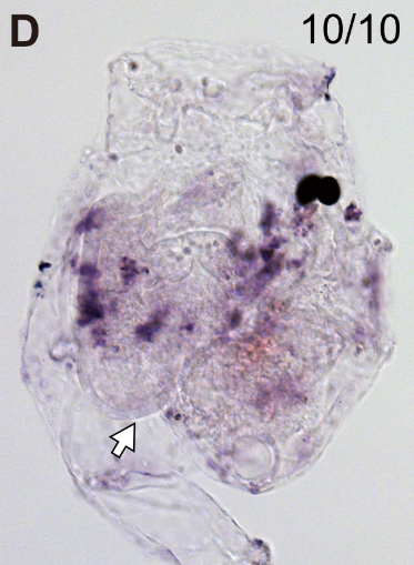
Lateral view of the head of a WT Ciona robusta swimming larva probed for Hox1 expression.
Expression of Hox1 in the endoderm marks presumptive posterior pharynx (white arrows).

Stained molecule KH2012:KH.L171.16 (HOXA1; HOXB1; HOXD1)
Stained region(s)

C. robusta formely Ciona int. type A, Stage 27 (swimming larva I)

Read more…

Lateral view of the head of a Ciona robusta swimming larva probed for Hox1 expression and electroporated with Titf>Otx-TALEN. This overexpression construct is used to target the Otx locus in the endodermal tissue.
Expression of Hox1 in the endoderm marks presumptive posterior pharynx (white arrows) and was not affected by Otx KO.

Stained molecule KH2012:KH.L171.16 (HOXA1; HOXB1; HOXD1)
Stained region(s)

C. robusta formely Ciona int. type A, Stage 37 (Hotta stage 37)

Read more…

WT Ciona robusta 3 days post fertilization juvenile probed for Hox1 expression.
Hox1 is expressed in the posterior tip of the endostyle (black arrow).

Stained molecule KH2012:KH.L171.16 (HOXA1; HOXB1; HOXD1)
Stained region(s)

C. robusta formely Ciona int. type A, Stage 37 (Hotta stage 37)

Read more…

Ciona robusta 3 days post fertilization juvenile probed for Hox1 expression and electroporated with Titf>Raldh2-TALEN and Tnl>Raldh2-TALEN. These overexpression constructs are used to target the Hox1 locus in the endoderm and muscle respectively.
Expression of Hox1 in the posterior endostyle is lost (white arrow).
Scale bar: 50 µm.

Stained molecule KH2012:KH.L171.16 (HOXA1; HOXB1; HOXD1)
Stained region(s)

C. robusta formely Ciona int. type A, Stage 39 (Hotta stage 39)

Read more…

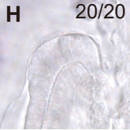
Expression was detected in the posterior endostyle.

Stained molecule KH2012:KH.L171.16 (HOXA1; HOXB1; HOXD1)
Stained region(s)

C. robusta formely Ciona int. type A, Stage 39 (Hotta stage 39)

Read more…

Whole-mount in situ hybridization for Hox1 in a 6 days post fertilization WT Ciona robusta juvenile.
Hox1 is expressed in the posterior endostyle (black arrows).
Scale bar: 50 µm.
The second image is a magnified anterior tip of the endostyle of another embryo.

Stained molecule KH2012:KH.L171.16 (HOXA1; HOXB1; HOXD1)
Stained region(s)

C. robusta formely Ciona int. type A, Stage 39 (Hotta stage 39)

Read more…

6 days post fertilization Ciona robusta juvenile probed for Hox1 expression and electroporated with Titf>Hox1-TALEN. This overexpression construct is used to target the Hox1 locus in the endodermal tissue.
Hox1 expression in the posterior endostyle is lost (white arrows).

Stained molecule KH2012:KH.L171.16 (HOXA1; HOXB1; HOXD1)
Stained region(s) No expression -

C. robusta formely Ciona int. type A, Stage 39 (Hotta stage 39)

Read more…

6 days post fertilization Ciona robusta juvenile probed for Hox1 expression and electroporated with Titf>Otx-TALEN. This overexpression construct is used to target the Otx locus in the endodermal tissue.
Hox1 is expressed in the posterior endostyle, as in WT conditions (black arrows), but also in the anterior endostyle (red arrows).
The second image is the magnified posterior tip of the endostyle of another embryo.

Stained molecule KH2012:KH.L171.16 (HOXA1; HOXB1; HOXD1)
Stained region(s)

C. robusta formely Ciona int. type A, Stage 39 (Hotta stage 39)

Read more…

6 days post fertilization Ciona robusta juvenile probed for Hox1 expression and co-electroporated with Titf>Otx-TALEN and Titf>Hox1-TALEN. These overexpression constructs are used to target Otx and Hox1 loci in the endodermal tissue.
Hox1 expression in the posterior endostyle is lost (white arrows).

Stained molecule KH2012:KH.L171.16 (HOXA1; HOXB1; HOXD1)
Stained region(s)

C. robusta formely Ciona int. type A, Stage 39 (Hotta stage 39)

Read more…

6 days post fertilization Ciona robusta juvenile probed for Hox1 expression and electroporated with Titf>Otx. This construct is used to overexpress Otx in the endodermal tissue.
Hox1 expression in the posterior endostyle is lost (white arrows).

Stained molecule KH2012:KH.L171.16 (HOXA1; HOXB1; HOXD1)
Stained region(s)

C. robusta formely Ciona int. type A, Stage 48 (young adult)

Read more…

Expression signals were assessed by whole-mount in situ hybridization (WISH) and the NBT/BCIP coloring system under the stereomicroscope.
A. Representation of a 2– to 3-week-old Ciona juvenile, left side view (anterior, as atrial siphon, bs branchial sac, d dorsal, epc epicardium, endostyle, esp esophagus, int intestine, nc neural complex, os oral siphon, rpb retropharyngeal band, p posterior, pj protruding structures of the pharyngeal esophagus junction, st stomach, stg stigma, v ventral).
B. Hox1 expression in the posterior endostyle, retropharyngeal band, epicardium and protruding structures of the pharyngeal esophagus junction. The blue stain (asterisk) in the stomach is a nonspecific stain of the intestinal inclusions. Bar 500 μm
“In 2– to 3-week-old juvenile specimens, the differential expression patterns of all genes in the ascidian Hox cluster were detected via a stereo microscope”

Stained molecule KH2012:KH.L171.16 (HOXA1; HOXB1; HOXD1)
Stained region(s)

C. robusta formely Ciona int. type A, Stage 48 (young adult)

Read more…

The blue stains (asterisks) in the stomach are nonspecific stains of intestinal inclusions (en endostyle, esp esophagus, int intestine, st stomach). Bars 100 μm
In the protruding structures of the pharyngeal esophagus junction (white arrowheads ventral side, gray arrowheads dorsal side, black arrowheads distal side), expression of Hox1 is observed.
“In the endostyle, Hox1 is expressed. For each gene, expression in the posterior region is stronger than the anterior region and the strongest signal was detected in the posterior terminus (white arrowheads).”

Stained molecule KH2012:KH.L171.16 (HOXA1; HOXB1; HOXD1)
Stained region(s)

C. robusta formely Ciona int. type A, Stage 48 (young adult)

Read more…

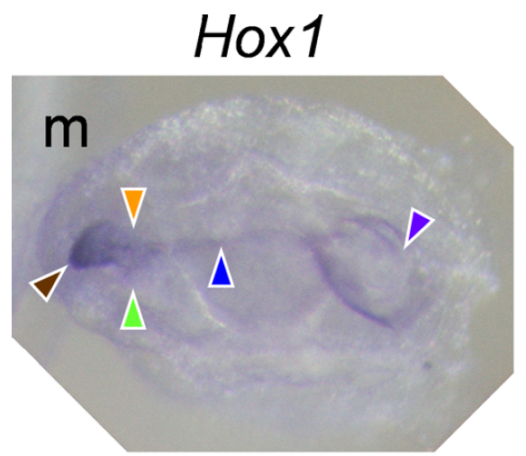
Expression domains are indicated by colored arrowheads: brown endostyle, purple esophagus, blue retropharyngeal band, green right epicardium, orange left side of left epicardium, red right side of left epicardium. Bar 100 μm
Expression was assessed from the left (a–d), right (e–h), anteroventral (i–l) and opposite views of the oral siphon (m–p). Expression was also assessed by using frontally sectioned specimens (q–t).
“Expression of Hox1 was restricted near the retropharyngeal band and was slightly expanded lateral to the epicardium (Fig. 3a, e, i, m, q).”

Stained molecule KH2012:KH.L171.16 (HOXA1; HOXB1; HOXD1)
Stained region(s)

C. robusta formely Ciona int. type A, Stage 48 (young adult)

Read more…

a Expression of Hox1 is observed in the middle to posterior part of the cerebral ganglion. Bar 20 μm.

Stained molecule KH2012:KH.L171.16 (HOXA1; HOXB1; HOXD1)
Stained region(s)