Login
Help

GENE CARD

Submit your Data

EST count

27 results

Library Name Percentage Expression
Ciona intestinalis whole animal Ciona intestinalis whole animal 0 clone(s) / 0
Nori Satoh unpublished cDNA library Nori Satoh unpublished cDNA library 0 clone(s) / 5,577
Nori Satoh unpublished cDNA library, young adult Nori Satoh unpublished cDNA library, young adult 0 clone(s) / 31,366
Nori Satoh unpublished cDNA library, cleavage stage embryo Nori Satoh unpublished cDNA library, cleavage stage embryo 1 clone(s) / 15,579
Nori Satoh unpublished cDNA library, egg Nori Satoh unpublished cDNA library, egg 0 clone(s) / 31,100
Nori Satoh unpublished cDNA library, larva Nori Satoh unpublished cDNA library, larva 0 clone(s) / 26,704
Nori Satoh unpublished cDNA library, tailbud embryo Nori Satoh unpublished cDNA library, tailbud embryo 0 clone(s) / 26,680
directional larval cDNA library directional larval cDNA library 0 clone(s) / 39
Ascidian hemocytes cDNA library Ascidian hemocytes cDNA library 0 clone(s) / 0
K. Inaba unpublished cDNA library, testis K. Inaba unpublished cDNA library, testis 0 clone(s) / 5,468
Stratagene UniZAP whole-larva library Stratagene UniZAP whole-larva library 0 clone(s) / 0
Ciona intestinalis larva Ciona intestinalis larva 0 clone(s) / 0
Nori Satoh unpublished cDNA library, blood cells Nori Satoh unpublished cDNA library, blood cells 0 clone(s) / 29,579
Nori Satoh unpublished cDNA library, endostyle Nori Satoh unpublished cDNA library, endostyle 0 clone(s) / 2,556
Nori Satoh unpublished cDNA library, cleaving embryo Nori Satoh unpublished cDNA library, cleaving embryo 2 clone(s) / 16,939
Nori Satoh unpublished cDNA library, gastrula and neurula Nori Satoh unpublished cDNA library, gastrula and neurula 8 clone(s) / 25,258
Nori Satoh unpublished cDNA library, gonad Nori Satoh unpublished cDNA library, gonad 0 clone(s) / 16,936
Nori Satoh unpublished cDNA library, neural complex Nori Satoh unpublished cDNA library, neural complex 0 clone(s) / 10,463
Nori Satoh unpublished cDNA library, heart Nori Satoh unpublished cDNA library, heart 0 clone(s) / 13,243
Yutaka Satou unpublished cDNA library, adult digestive gland Yutaka Satou unpublished cDNA library, adult digestive gland 0 clone(s) / 17,765
Yutaka Satou unpublished cDNA library, embryo whole animal Yutaka Satou unpublished cDNA library, embryo whole animal 0 clone(s) / 17,872
Yutaka Satou unpublished cDNA library, mature adult whole animal Yutaka Satou unpublished cDNA library, mature adult whole animal 0 clone(s) / 107,314
Nori Satoh unpublished cDNA library, juvenile whole animal Nori Satoh unpublished cDNA library, juvenile whole animal 0 clone(s) / 24,372
Nori Satoh unpublished cDNA library, mature adult whole animal Nori Satoh unpublished cDNA library, mature adult whole animal 0 clone(s) / 17,126
Ciona intestinalis whole animal stage3 juvenile Ciona intestinalis whole animal stage3 juvenile 0 clone(s) / 3,798
Yutaka Satou unpublished library (cicx) Yutaka Satou unpublished library (cicx) 0 clone(s) / 2,031
Gateway compatible cien cDNA library, Ciona intestinalis mixed embryonic stages (Egg to Neurula) Gateway compatible cien cDNA library, Ciona intestinalis mixed embryonic stages (Egg to Neurula) 2 clone(s) / 188,431

RNA-Seq transcriptome profiles (by Experiment)

3 results

OVERVIEW

   Expression profile summary from all experiments in WT and mutant conditions:
FPKM & RPKM

Click here to see the RNA-Seq Pipeline Protocol.
You can show/hide lines in the charts by clicking on it in the legend part.
You can see more details by clicking on a point and then, on the link which will appear.

Be careful, FPKM & RPKM does not permit you to compare a gene across different conditions or experiments.

Download FPKM & RPKM data

OVERVIEW

   Expression profile summary from all experiments in WT and mutant conditions:
Relative Log Expression RLE

Be careful, RLE and 2RLE does not permit you to compare a gene across different experiments.

Download RLE Relative Log Expression data

RNA-Seq Experiment n°34135

Read more…

Oocytes of a single individual of Ciona intestinalis (Roscoff, France) were fertilized by a mixture of sperm of two individuals and development was followed microscopically. Embryos were snapfrozen in liquid nitrogen at the appropriate stage and kept at -80°C. Samples at stage 0, 8, 12, 15, 21, 26 were collected from the same fertilzation, while sample from stage 11 was independently collected from a different individual. Total RNA was prepared with the RNA II kit of Magerey Nagel and the quality was checked with the Bioanalyzer, all samples had a RIN value of higher than 9. Strand specific libraries were constructed and the sequencing (50PE) was performed with Illumina HiSeq 2000 technology (BGI, Hong Kong, China). Reads were aligned onto C.robusta genome and considered to reflect the expression of the C.robusta orthologous genes.

Assay platform Illumina HiSeq 2000
Layout Paired-end
Stranded Yes
Read length 49
Normalized data types
  • FPKM
  • RLE
Wild type - Replica 1
Territories whole embryo -
Biomaterial(s) C. robusta formely Ciona int. type A, Stage 0 (Unfertilized egg)
C. robusta formely Ciona int. type A, Stage 8 (64-cell)
C. robusta formely Ciona int. type A, Stage 11 (early gastrula)
C. robusta formely Ciona int. type A, Stage 12 (mid gastrula)
C. robusta formely Ciona int. type A, Stage 15 (mid neurula)
C. robusta formely Ciona int. type A, Stage 21 (mid tailbud I)
C. robusta formely Ciona int. type A, Stage 26 (hatching larva)
Wild type - Replica 2
Territories whole embryo -
Biomaterial(s) C. robusta formely Ciona int. type A, Stage 0 (Unfertilized egg)
C. robusta formely Ciona int. type A, Stage 8 (64-cell)
C. robusta formely Ciona int. type A, Stage 11 (early gastrula)
C. robusta formely Ciona int. type A, Stage 12 (mid gastrula)
C. robusta formely Ciona int. type A, Stage 15 (mid neurula)
C. robusta formely Ciona int. type A, Stage 21 (mid tailbud I)
C. robusta formely Ciona int. type A, Stage 26 (hatching larva)

RNA-Seq Experiment n°34138

Read more…

Ciona robusta adults were obtained from the National Bio-Resource Project for Ciona (NBRP, Japan). 50 unperturbed and Foxd-morphant embryos were collected at the 32-, 64-, and 112-cell stages. RNA was extracted using a Dynabeads mRNA DIRECT Purification Kit (Thermo Fischer Scientific) and libraries were made with an Ion Total RNA-Seq kit ver 2 (Thermo Fischer Scientific). The libraries were sequenced with an Ion PGM instrument (Thermo Fischer Scientific).

Assay platform Ion Torrent PGM
Layout Single-end
Stranded Yes
Read length 20-365
Normalized data types
  • RPKM
Wild type
Territories whole embryo -
Biomaterial(s) C. robusta formely Ciona int. type A, Stage 6a (early 32-cell)
C. robusta formely Ciona int. type A, Stage 8 (64-cell)
C. robusta formely Ciona int. type A, Stage 10 (112-cell)
Mutant
Deregulated molecule(s) KH2012:KH.C8.890 -
Territories whole embryo -
Biomaterial(s) C. robusta formely Ciona int. type A, Stage 6a (early 32-cell)
C. robusta formely Ciona int. type A, Stage 8 (64-cell)
C. robusta formely Ciona int. type A, Stage 10 (112-cell)

RNA-Seq Experiment n°34139

Read more…

The purpose of this project is to identify genes positively and negatively regulated by signaling molecules that belongs to the TGFbeta superfamily.

Assay platform Illumina HiSeq 2500
Layout Single-end
Stranded Yes
Read length 101
Normalized data types
  • RPKM
Wild type
Territories whole embryo -
Biomaterial(s) C. robusta formely Ciona int. type A, Stage 13 (late gastrula)
Mutant 1
Deregulated molecule(s) KH2012:KH.C4.125 -
Territories whole embryo -
Biomaterial(s) C. robusta formely Ciona int. type A, Stage 13 (late gastrula)
Mutant 2
Deregulated molecule(s) KH2012:KH.C2.573 -
Territories whole embryo -
Biomaterial(s) C. robusta formely Ciona int. type A, Stage 13 (late gastrula)

In Situ Experiment Data

113 results

Experiment data ( results)

C. robusta formely Ciona int. type A, Stage 1 (One cell)

Read more…

Conversion to Aniseed format of data from Yutaka Satou’s gene expression profiles of transcription factors and signalling molecule ISH screen (Development. 2004 131:4047-58).

Original annotation :
Gene name :Tbx6b/c/d.
Expression pattern : No signal was detected

Stained molecule KH2012:KH.S654.3 (TBX2; TBX4; TBX6)
Stained region(s) No expression -

C. robusta formely Ciona int. type A, Stage 2 (2-cell)

Read more…

WT Ciona robusta 2-cell stage embryo (anterior to the left) probed for Tbx6-r.b expression.
No Tbx6-r.b expression is observed.

Stained molecule KH2012:KH.S654.3 (TBX2; TBX4; TBX6)
Stained region(s) No expression -

C. robusta formely Ciona int. type A, Stage 2 (2-cell)

Read more…

Ciona robusta 2-cell stage embryo (anterior to the left) probed for Tbx6-r.b expression and simultaneously injected with MOs targeting Hes.a, Pem and Ccnb3.
No Tbx6-r.b expression is observed.

Stained molecule KH2012:KH.S654.3 (TBX2; TBX4; TBX6)
Stained region(s) No expression -

C. robusta formely Ciona int. type A, Stage 3 (4-cell)

Read more…

WT Ciona robusta 4-cell stage embryo (anterior to the left) probed for Tbx6-r.b expression.
No Tbx6-r.b expression is observed.

Stained molecule KH2012:KH.S654.3 (TBX2; TBX4; TBX6)
Stained region(s) No expression -

C. robusta formely Ciona int. type A, Stage 3 (4-cell)

Read more…

Ciona robusta 4-cell stage embryo (anterior to the left) probed for Tbx6-r.b expression and simultaneously injected with MOs targeting Hes.a, Pem and Ccnb3.
No Tbx6-r.b expression is observed.

Stained molecule KH2012:KH.S654.3 (TBX2; TBX4; TBX6)
Stained region(s) No expression -

C. robusta formely Ciona int. type A, Stage 4 (8-cell)

Read more…

WT Ciona robusta 8-cell stage embryo (anterior to the left) probed for Tbx6-r.b expression.
No Tbx6-r.b expression is observed.

Stained molecule KH2012:KH.S654.3 (TBX2; TBX4; TBX6)
Stained region(s) No expression -

C. robusta formely Ciona int. type A, Stage 4 (8-cell)

Read more…

Ciona robusta 8-cell stage embryo (anterior to the left) probed for Tbx6-r.b expression and simultaneously injected with MOs targeting Hes.a, Pem and Ccnb3.
No Tbx6-r.b expression is observed.

Stained molecule KH2012:KH.S654.3 (TBX2; TBX4; TBX6)
Stained region(s) No expression -

C. robusta formely Ciona int. type A, Stage 4 (8-cell)

Read more…

Conversion to Aniseed format of data from Yutaka Satou’s gene expression profiles of transcription factors and signalling molecule ISH screen (Development. 2004 131:4047-58).

Original annotation :
Gene name :Tbx6b/c/d.
Expression pattern : no expression.

Stained molecule KH2012:KH.S654.3 (TBX2; TBX4; TBX6)
Stained region(s) No expression -

C. robusta formely Ciona int. type A, Stage 5a (early 16-cell)

Read more…

Vegetal view of a WT Ciona robusta 16-cell stage embryo probed for Tbx6-r.b expression (and injected with iin vitro-transcribed GFP:H2B control mRNA).
Tbx6-r.b expression is observed in B5.1 blastomer pair.

Stained molecule KH2012:KH.S654.3 (TBX2; TBX4; TBX6)
Stained region(s) B5.1 cell pair -

C. robusta formely Ciona int. type A, Stage 5a (early 16-cell)

Read more…

Vegetal view of a Ciona robusta 16-cell stage embryo probed for Tbx6-r.b expression and injected with Ccnb3 mRNA.
No Tbx6-r.b expression is observed.
Original annotation: Ccnb3 overexpression resulted in the failure to detect any expression of early zygotic genes and a loss of cell adhesion.

Stained molecule KH2012:KH.S654.3 (TBX2; TBX4; TBX6)
Stained region(s) No expression -

C. robusta formely Ciona int. type A, Stage 5a (early 16-cell)

Read more…

Vegetal view of a Ciona robusta 16-cell stage embryo probed for Tbx6-r.b expression and injected with Ccnb3 mRNA with mutations in the cdk-binding domain (Ccnb3 AAA mRNA).
Tbx6-r.b expression is observed in B5.1 blastomer pair, as in the WT control.
Original annotation: Expression was not lost in embryos injected with a mutant form of the Ccnb3 mRNA harboring inactivating amino acid substitutions in the cdk-binding site (Ccnb3 AAA).

Stained molecule KH2012:KH.S654.3 (TBX2; TBX4; TBX6)
Stained region(s) B5.1 cell pair -

C. robusta formely Ciona int. type A, Stage 5a (early 16-cell)

Read more…

Vegetal view of a Ciona robusta 16-cell stage embryo probed for Tbx6-r.b expression and injected with Ccnb1 mRNA.
Tbx6-r.b expression is observed in B5.1 blastomer pair, as in the WT control.
Original annotation: Expression was not lost in embryos injected with Ccnb1 mRNA.

Stained molecule KH2012:KH.S654.3 (TBX2; TBX4; TBX6)
Stained region(s) B5.1 cell pair -

C. robusta formely Ciona int. type A, Stage 5a (early 16-cell)

Read more…

Vegetal view of a 16-cell stage embryo probed for Ci-Tbx6b expression. Cells where mRNA is first detected are indicated by arrowheads.

Stained molecule KH2012:KH.S654.3 (TBX2; TBX4; TBX6)
Stained region(s) B5.1 cell pair -

C. robusta formely Ciona int. type A, Stage 5a (early 16-cell)

Read more…

Vegetal view of a 16-cell stage 5-aza-dC- treated embryo probed for Ci-Tbx6b expression.

Original annotation:
Appearance of the Ci-Tbx6b mRNAs was retarded.

Stained molecule KH2012:KH.S654.3 (TBX2; TBX4; TBX6)
Stained region(s) No expression -

C. robusta formely Ciona int. type A, Stage 5a (early 16-cell)

Read more…

Conversion to Aniseed format of data from Yutaka Satou's gene expression profiles of transcription factors and signalling molecule ISH screen (Development. 2004 131:4047-58).

Original annotation :
Gene name :Tbx6b/c/d.
Expression pattern : B5.1

Stained molecule KH2012:KH.S654.3 (TBX2; TBX4; TBX6)
Stained region(s) B5.1 cell pair -

C. robusta formely Ciona int. type A, Stage 5a (early 16-cell)

Read more…

Vegetal pole view of a 16-cell stage embryo probed with Ci-Tbx6b expression.

Stained molecule KH2012:KH.S654.3 (TBX2; TBX4; TBX6)
Stained region(s) B5.1 cell pair -

C. robusta formely Ciona int. type A, Stage 5a (early 16-cell)

Read more…

Vegetal pole view of a 16-cell stage embryo probed with Ci-Tbx6b expression. Injection of Ci-macho1 MO prevents the expression of Ci-Tbx6b contrary to the control where Ci-Tbx6b is expressed in the B5.1 cell pair.

Stained molecule KH2012:KH.S654.3 (TBX2; TBX4; TBX6)
Stained region(s) No expression -

C. robusta formely Ciona int. type A, Stage 5a (early 16-cell)

Read more…

Vegetal pole view of a 16-cell stage embryo probed with Ci-Tbx6b expression. Injection of Ci-macho1 MO and Ci-ZicL MO prevents the expression of Ci-Tbx6b contrary to the control where Ci-Tbx6b is expressed in the B5.1 cell pair.

Stained molecule KH2012:KH.S654.3 (TBX2; TBX4; TBX6)
Stained region(s) No expression -

C. robusta formely Ciona int. type A, Stage 5a (early 16-cell)

Read more…

Animal and vegetal views of a 16-cell stage embryo probed for Tbx6.b expression.
Original annotation: As observed in normal embryos, in embryos injected with the control MO, Tbx6.b was expressed only in the PVD.

Stained molecule KH2012:KH.S654.3 (TBX2; TBX4; TBX6)
Stained region(s) B5.1 cell pair -

C. robusta formely Ciona int. type A, Stage 5a (early 16-cell)

Read more…

Animal and vegetal views of a 16-cell stage embryo probed for Tbx6.b expression, injected with Gata.a-MO.
Original annotation: The Tbx6.b expression was unaffected in Gata.a morphants.

Stained molecule KH2012:KH.S654.3 (TBX2; TBX4; TBX6)
Stained region(s) B5.1 cell pair -

C. robusta formely Ciona int. type A, Stage 5a (early 16-cell)

Read more…

Animal and vegetal views of a 16-cell stage embryo probed for Tbx6.b expression, injected with beta-catenin-MO.
Original annotation: The Tbx6.b expression was lost in β-catenin morphants.

Stained molecule KH2012:KH.S654.3 (TBX2; TBX4; TBX6)
Stained region(s) No expression -

C. robusta formely Ciona int. type A, Stage 5a (early 16-cell)

Read more…

Animal and vegetal views of a 16-cell stage embryo probed for Tbx6.b expression, injected with Zic-r.a MO.
Original annotation: The Tbx6.b expression was lost in Zic-r.a morphants.

Stained molecule KH2012:KH.S654.3 (TBX2; TBX4; TBX6)
Stained region(s) No expression -

C. robusta formely Ciona int. type A, Stage 5a (early 16-cell)

Read more…

Expression of Ci-Tbx6b was detected by whole-mount in situ hybridization. Vegetal view of a 16-cell stage embryo.

Stained molecule KH2012:KH.S654.3 (TBX2; TBX4; TBX6)
Stained region(s) B5.1 cell pair -

C. robusta formely Ciona int. type A, Stage 5a (early 16-cell)

Read more…

Illustrations on the left depict the constructs. The numbers indicate the relative nucleotide positions from the transcription start site of Tbx6.b. Mutated Tcf7-binding sites are indicated by X. Graphs show the percentage of blastomeres expressing the reporter in the anterior vegetal blastomeres, in the posterior vegetal blastomeres, and in the animal blastomeres. Note that not all cells or embryos could express the reporter because of mosaic incorporation of the electroporated plasmid.
Images showing Gfp expression in embryos electroporated with the fourth and last constructs shown in (A). Scale bar, 100 μm.
"Our analyses using a series of deletion reporter constructs identified the −189 bp upstream of Tbx6.b as a region sufficient to drive reporter expression in the PVD at the 16-cell stage"

Stained molecule KH2012:KH.S654.3 (TBX2; TBX4; TBX6)
Stained region(s) B5.1 cell pair -

C. robusta formely Ciona int. type A, Stage 5a (early 16-cell)

Read more…

Illustrations on the left depict the constructs. The numbers indicate the relative nucleotide positions from the transcription start site of Tbx6.b. Mutated Tcf7-binding sites are indicated by X. Graphs show the percentage of blastomeres expressing the reporter in the anterior vegetal blastomeres, in the posterior vegetal blastomeres, and in the animal blastomeres. Note that not all cells or embryos could express the reporter because of mosaic incorporation of the electroporated plasmid.

Stained molecule KH2012:KH.S654.3 (TBX2; TBX4; TBX6)
Stained region(s) No expression -

C. robusta formely Ciona int. type A, Stage 5a (early 16-cell)

Read more…

Illustrations on the left depict the constructs. The numbers indicate the relative nucleotide positions from the transcription start site of Tbx6.b. Mutated Tcf7-binding sites are indicated by X. Graphs show the percentage of blastomeres expressing the reporter in the anterior vegetal blastomeres, in the posterior vegetal blastomeres, and in the animal blastomeres. Note that not all cells or embryos could express the reporter because of mosaic incorporation of the electroporated plasmid.
Images showing Gfp expression in embryos electroporated with the fourth and last constructs shown in (A). Scale bar, 100 μm.
"When two putative Tcf7-binding sites were mutated, the expression of the reporter was completely abolished"

Stained molecule KH2012:KH.S654.3 (TBX2; TBX4; TBX6)
Stained region(s) No expression -

C. robusta formely Ciona int. type A, Stage 5a (early 16-cell)

Read more…

Illustrations on the left depict the constructs. The numbers indicate the relative nucleotide positions from the transcription start site of Tbx6.b. Mutated Tcf7-binding sites are indicated by X. Graphs show the percentage of blastomeres expressing the reporter in the anterior vegetal blastomeres, in the posterior vegetal blastomeres, and in the animal blastomeres. Note that not all cells or embryos could express the reporter because of mosaic incorporation of the electroporated plasmid.

Stained molecule KH2012:KH.S654.3 (TBX2; TBX4; TBX6)
Stained region(s) B5.1 cell pair -

C. robusta formely Ciona int. type A, Stage 5a (early 16-cell)

Read more…

First picture
Four repeats of the critical upstream sequence of Tbx6.b with or without mutations were ligated to the Brachyury basal promoter. Graphs show the percentage of blastomeres expressing the reporter among the anterior vegetal blastomeres and among the posterior vegetal blastomeres. A graph showing the percentage in the animal hemisphere is not shown, because no embryos expressed the reporter in the animal hemisphere. Note that not all cells or embryos could express the reporter because of mosaic incorporation of the electroporated plasmid.
Second picture
Images showing the reporter gene expression in embryos electroporated with the construct shown in the first image.
“While the basal promoter alone cannot drive reporter expression, the fusion construct was specifically expressed in the PVD. Therefore, these sequences contained sufficient elements to drive the reporter expression in the PVD.”

Stained molecule KH2012:KH.S654.3 (TBX2; TBX4; TBX6)
Stained region(s) B5.1 cell pair -

C. robusta formely Ciona int. type A, Stage 5a (early 16-cell)

Read more…

Four repeats of the critical upstream sequence of Tbx6.b with or without mutations were ligated to the Brachyury basal promoter. Graphs show the percentage of blastomeres expressing the reporter among the anterior vegetal blastomeres and among the posterior vegetal blastomeres. A graph showing the percentage in the animal hemisphere is not shown, because no embryos expressed the reporter in the animal hemisphere. Note that not all cells or embryos could express the reporter because of mosaic incorporation of the electroporated plasmid.
Second picture
Images showing the reporter gene expression in embryos electroporated with the construct shown in the first image.
“While the basal promoter alone cannot drive reporter expression, the fusion construct was specifically expressed in the PVD. Therefore, these sequences contained sufficient elements to drive the reporter expression in the PVD.”
“The mutant construct μC induced ectopic expression of the reporter in the AVD. Therefore, the region mutated in μC (region C) is likely to repress expression in the AVD”

Stained molecule KH2012:KH.S654.3 (TBX2; TBX4; TBX6)
Stained region(s) A5.1 cell pair - A5.2 cell pair - B5.1 cell pair -

C. robusta formely Ciona int. type A, Stage 5a (early 16-cell)

Read more…

Four repeats of the critical upstream sequence of Tbx6.b with or without mutations were ligated to the Brachyury basal promoter. Graphs show the percentage of blastomeres expressing the reporter among the anterior vegetal blastomeres and among the posterior vegetal blastomeres. A graph showing the percentage in the animal hemisphere is not shown, because no embryos expressed the reporter in the animal hemisphere. Note that not all cells or embryos could express the reporter because of mosaic incorporation of the electroporated plasmid.
“While the basal promoter alone cannot drive reporter expression, the fusion construct was specifically expressed in the PVD. Therefore, these sequences contained sufficient elements to drive the reporter expression in the PVD.”

Stained molecule KH2012:KH.S654.3 (TBX2; TBX4; TBX6)
Stained region(s) B5.1 cell pair -

C. robusta formely Ciona int. type A, Stage 5a (early 16-cell)

Read more…

Four repeats of the critical upstream sequence of Tbx6.b with or without mutations were ligated to the Brachyury basal promoter. Graphs show the percentage of blastomeres expressing the reporter among the anterior vegetal blastomeres and among the posterior vegetal blastomeres. A graph showing the percentage in the animal hemisphere is not shown, because no embryos expressed the reporter in the animal hemisphere. Note that not all cells or embryos could express the reporter because of mosaic incorporation of the electroporated plasmid.
“While the basal promoter alone cannot drive reporter expression, the fusion construct was specifically expressed in the PVD. Therefore, these sequences contained sufficient elements to drive the reporter expression in the PVD.”

Stained molecule KH2012:KH.S654.3 (TBX2; TBX4; TBX6)
Stained region(s) B5.1 cell pair -

C. robusta formely Ciona int. type A, Stage 5a (early 16-cell)

Read more…

Illustrations on the left depict the constructs. A potential repressive element in the upstream sequence of Tbx6.b was inserted into the upstream sequences of Fgf9/16/20. Graphs on the right show the percentage of blastomeres expressing the reporter gene in the anterior vegetal blastomeres and in the posterior vegetal blastomeres. A graph showing the percentage in the animal hemisphere is not shown, because no embryos expressed the reporter in the animal hemisphere.
"With this insertion, the construct rarely drove reporter expression in the AVD"
"Although the result does not necessarily indicate that the 22 bp region is the only repressive element in the upstream region of Tbx6.b, it showed that this 22 bp region has a repressive activity."

Stained molecule KH2012:KH.C2.125 (FGF16; FGF20; FGF9)
Stained region(s) B5.1 cell pair -

C. robusta formely Ciona int. type A, Stage 5a (early 16-cell)

Read more…

Stained molecule KH2012:KH.C8.396 (FOXD1; FOXD3; FOXD4)
Stained region(s) A5.1 cell pair - A5.2 cell pair - B5.1 cell pair -

C. robusta formely Ciona int. type A, Stage 5a (early 16-cell)

Read more…

Constructs depicted in the illustrations on the left were injected with or without an MO against Zic-r.a. The green boxes indicate the Gfp reporter gene and SV40 polyadenylation signal. Graphs on the right show the percentage of blastomeres expressing the reporter gene in the anterior vegetal blastomeres and in the posterior vegetal blastomeres.

Stained molecule KH2012:KH.S654.3 (TBX2; TBX4; TBX6)
Stained region(s) B5.1 cell pair -

C. robusta formely Ciona int. type A, Stage 5a (early 16-cell)

Read more…

First picture
Constructs depicted in the illustrations on the left were injected with or without an MO against Zic-r.a. Graphs on the right show the percentage of blastomeres expressing the reporter gene in the anterior vegetal blastomeres and in the posterior vegetal blastomeres.
Second picture
Photographs of embryo injected with the Fgf9/16/20 reporter construct with the repressive element of Tbx6.b. Scale bar, 100 μm.

Stained molecule KH2012:KH.C2.125 (FGF16; FGF20; FGF9)
Stained region(s) A5.1 cell pair - A5.2 cell pair - B5.1 cell pair -

C. robusta formely Ciona int. type A, Stage 6b (late 32-cell)

Read more…

Vegetal view of a 32-cell stage embryo probed for Ci-Tbx6b expression.

Stained molecule KH2012:KH.S654.3 (TBX2; TBX4; TBX6)
Stained region(s) B6.1 cell pair - B6.4 cell pair -

C. robusta formely Ciona int. type A, Stage 6b (late 32-cell)

Read more…

Vegetal view of a 32-cell stage 5-aza-dC- treated embryo probed for Ci-Tbx6b expression.

Original annotation:
Appearance of the Ci-Tbx6b mRNAs was retarded.

Stained molecule KH2012:KH.S654.3 (TBX2; TBX4; TBX6)
Stained region(s) No expression -

C. robusta formely Ciona int. type A, Stage 6b (late 32-cell)

Read more…

Conversion to Aniseed format of data from Yutaka Satou's gene expression profiles of transcription factors and signalling molecule ISH screen (Development. 2004 131:4047-58).

Original annotation :
Gene name :Tbx6b/c/d.
Expression pattern : B6.2, B6.4
Comment : The expression in the B6.2 is dispersed within cytoplasm and looked weak.

Stained molecule KH2012:KH.S654.3 (TBX2; TBX4; TBX6)
Stained region(s) B6.2 cell pair - B6.4 cell pair -

C. robusta formely Ciona int. type A, Stage 6b (late 32-cell)

Read more…

Vegetal view of a late 32-cell stage embryo.Tbx6b expression was detected in B6.1, B6.2, B6.4 blastomeres (signal in B6.1 and B6.2 is scarcely visible).

Stained molecule KH2012:KH.S654.3 (TBX2; TBX4; TBX6)
Stained region(s) B6.1 cell pair - B6.2 cell pair - B6.4 cell pair -

C. robusta formely Ciona int. type A, Stage 6b (late 32-cell)

Read more…

Vegetal view of a late 32-cell stage embryo. In ci-macho1-overexpressing embryos, transcript of Tbx6b gene was ectopically detected in vegetal blastomeres.

Stained molecule KH2012:KH.S654.3 (TBX2; TBX4; TBX6)
Stained region(s) A line - B line -

C. robusta formely Ciona int. type A, Stage 6b (late 32-cell)

Read more…

Vegatal view of a late 32-cell stage embryo. Effects of functional suppression of Ci-macho1 revealed by whole-mount in situ hybridization. Tbx6b expression disappeared in Ci-macho1-suppressed embryos.

Stained molecule KH2012:KH.S654.3 (TBX2; TBX4; TBX6)
Stained region(s) No expression -

C. robusta formely Ciona int. type A, Stage 6b (late 32-cell)

Read more…

Vegetal pole view of a late 32-cell stage embryo probed with Ci-Tbx6b expression.

Stained molecule KH2012:KH.S654.3 (TBX2; TBX4; TBX6)
Stained region(s) B6.2 cell pair - B6.4 cell pair -

C. robusta formely Ciona int. type A, Stage 6b (late 32-cell)

Read more…

Vegetal pole view of a late 32-cell stage embryo probed with Ci-Tbx6b expression. Injection of Ci-macho1 prevents the expression of Ci-Tbx6b contrary to the control where Ci-Tbx6b is expressed in the B6.2 and B6.4 cell pairs.

Stained molecule KH2012:KH.S654.3 (TBX2; TBX4; TBX6)
Stained region(s) No expression -

C. robusta formely Ciona int. type A, Stage 6b (late 32-cell)

Read more…

Vegetal pole view of an early 32-cell stage embryo probed with Ci-Tbx6b expression. Injection of Ci-macho1 and Ci-ZicL MO prevents the expression of Ci-Tbx6b contrary to the control where Ci-Tbx6b is expressed in the B6.2 and B6.4 cell pairs.

Stained molecule KH2012:KH.S654.3 (TBX2; TBX4; TBX6)
Stained region(s) No expression -

C. robusta formely Ciona int. type A, Stage 6b (late 32-cell)

Read more…

Expression of Ci-Tbx6b was detected by whole-mount in situ hybridization. Vegetal view of a late 32-cell stage embryo.

Stained molecule KH2012:KH.S654.3 (TBX2; TBX4; TBX6)
Stained region(s) B6.4 cell pair -

C. robusta formely Ciona int. type A, Stage 6b (late 32-cell)

Read more…

32-cell stage embryos electroporated with the 2.4 kb wild-type Ci-Tbx6b CRM, region upstream of Ci-Tbx6b from -2339bp to -13bp.
The first picture is a schematic representation of the electroporated region. Green boxes represent putative Ci-Macho1-binding sites; black arrows indicate their orientations. The orange rectangle depicts the proximal region of the 2.4 kb fragment.
The percentages of transgenic embryos displaying the patterns shown, and the total number of embryos scored, are indicated at the bottom of each panel.

Original Annotation : A strong hybridization signal was detected in the B6.4 pair of muscle precursors.

Stained molecule KH2012:KH.S654.3 (TBX2; TBX4; TBX6)
Stained region(s) B6.4 - B6.4* -

C. robusta formely Ciona int. type A, Stage 6b (late 32-cell)

Read more…

32-cell stage embryos electroporated with the 2.4 kb CRM with mutations in all Ci-Macho1-binding sites.
The first picture is a schematic representation of the electroporated region. Green boxes represent putative Ci-Macho1-binding sites; black arrows indicate their orientations. The orange rectangle depicts the proximal region of the 2.4 kb fragment.
The percentages of transgenic embryos displaying the patterns shown, and the total number of embryos scored, are indicated at the bottom of each panel.

Original Annotation : No expression of the reporter at the 32-cell stage.

Stained molecule KH2012:KH.S654.3 (TBX2; TBX4; TBX6)
Stained region(s) No expression -

C. robusta formely Ciona int. type A, Stage 6b (late 32-cell)

Read more…

32-cell stage embryos electroporated with the 862 bp Ci-Tbx6b CRM, which does not contain the Ci-Macho1-binding sites.
The first picture is a schematic representation of the electroporated region. The orange rectangle depicts the proximal region of the 2.4 kb fragment.
The percentages of transgenic embryos displaying the patterns shown, and the total number of embryos scored, are indicated at the bottom of each panel.

Original Annotation : No detectable signal at the 32-cell stage.

Stained molecule KH2012:KH.S654.3 (TBX2; TBX4; TBX6)
Stained region(s) No expression -

C. robusta formely Ciona int. type A, Stage 6b (late 32-cell)

Read more…

32-cell stage embryos electroporated with the fragment 266bp Ci-Tbx6b CRM containing the region upstream of Ci-Tbx6b from -145bp to -13bp upstream of basal promoter of Ci-Tbx6b.
The first picture is a schematic representation of the electroporated region.
The 266 bp proximal region of the Ci-Tbx6b CRM contains putative binding sites for CREB (blue diamond), T-box (red oval) and bHLH ("AC"-core E-box; pink triangle) transcription factors.
The percentages of transgenic embryos displaying the patterns shown, and the total number of embryos scored, are indicated at the bottom of each panel.

Original Annotation : The majority of the embryos electroporated with the 266 bp construct did not show staining at the 32-cell stage.

Stained molecule KH2012:KH.S654.3 (TBX2; TBX4; TBX6)
Stained region(s) No expression -

C. robusta formely Ciona int. type A, Stage 6b (late 32-cell)

Read more…

Embryos electroporated with either the distal muscle CRM and fixed at the 32-cell stage.
The distal region contains two E-boxes (pink triangles).
The percentages of transgenic embryos displaying the patterns shown, and the total number of embryos scored, are indicated at the bottom of each panel.

No reporter expression was detected.

Stained molecule KH2012:KH.S654.3 (TBX2; TBX4; TBX6)
Stained region(s) No expression -

C. robusta formely Ciona int. type A, Stage 8 (64-cell)

Read more…

Vegetal view of a 64-cell stage wild type embryo, showing expression of Ci-Tbx6b.

According to the picture, Ci-Tbx6b was expressed in B7.7, B7.5, B7.4 and B.8 cell pair.

Stained molecule KH2012:KH.S654.3 (TBX2; TBX4; TBX6)
Stained region(s) B7.4 cell pair - B7.5 cell pair - B7.7 cell pair - B7.8 cell pair -

C. robusta formely Ciona int. type A, Stage 8 (64-cell)

Read more…

Vegetal view of a 64-cell stage embryos, showing expression of Ci-Tbx6b following the injection of Ci-Rga-MO.

Ci-Tbx6b was still expressed in B7.7, B7.5, B7.4 and B.8 cell pair for 60% of embryos.

Stained molecule KH2012:KH.S654.3 (TBX2; TBX4; TBX6)
Stained region(s) B7.4 cell pair - B7.5 cell pair - B7.7 cell pair - B7.8 cell pair -

C. robusta formely Ciona int. type A, Stage 8 (64-cell)

Read more…

Conversion to Aniseed format of data from Yutaka Satou's gene expression profiles of transcription factors and signalling molecule ISH screen (Development. 2004 131:4047-58).

Original annotation :
Gene name :Tbx6b/c/d.
Expression pattern : B7.3, B7.4, B7.5, B7.7, B7.8.
Comment : The expression in the B7.3 and B7.4 is dispersed within cytoplasm and looked weak.

Stained molecule KH2012:KH.S654.3 (TBX2; TBX4; TBX6)
Stained region(s) B7.3 cell pair - B7.4 cell pair - B7.5 cell pair - B7.7 cell pair - B7.8 cell pair -

C. robusta formely Ciona int. type A, Stage 8 (64-cell)

Read more…

Vegetal pole view of a 64-cell stage embryo probed with Ci-Tbx6b expression.

Stained molecule KH2012:KH.S654.3 (TBX2; TBX4; TBX6)
Stained region(s) B7.4 cell pair - B7.5 cell pair - B7.7 cell pair - B7.8 cell pair -

C. robusta formely Ciona int. type A, Stage 8 (64-cell)

Read more…

Vegetal pole view of a 64-cell stage embryo probed with Ci-Tbx6b. Injection of Ci-macho1 MO seems to prevent the expression of Ci-Tbx6b in B7.4 and B7.5 cell pairs contrary to the control where Ci-Tbx6b is expressed in the B7.4, B7.5, B7.7 and B7.8 cell pairs.

Stained molecule KH2012:KH.S654.3 (TBX2; TBX4; TBX6)
Stained region(s) B7.7 cell pair -

C. robusta formely Ciona int. type A, Stage 8 (64-cell)

Read more…

Vegetal pole view of a 64-cell stage embryo probed with Ci-Tbx6b expression. Injection of Ci-macho1 MO and Ci-ZicL MO prevents the expression of Ci-Tbx6b contrary to the control where Ci-Tbx6b is expressed in the B7.4, B7.5, B7.7 and B7.8 cell pairs.

Stained molecule KH2012:KH.S654.3 (TBX2; TBX4; TBX6)
Stained region(s) No expression -

C. robusta formely Ciona int. type A, Stage 8 (64-cell)

Read more…

Expression of Ci-Tbx6b was detected by whole-mount in situ hybridization. Vegetal view of a 64-cell stage embryo.

Stained molecule KH2012:KH.S654.3 (TBX2; TBX4; TBX6)
Stained region(s) B7.4 cell pair - B7.5 cell pair - B7.7 cell pair - B7.8 cell pair -

C. robusta formely Ciona int. type A, Stage 10 (112-cell)

Read more…

Vegetal view of 110-cell stage wild type embryo probed for Tbx6b expression.

Stained molecule KH2012:KH.S654.3 (TBX2; TBX4; TBX6)
Stained region(s) B7.5 cell pair - B8.15 cell pair - B8.16 cell pair - B8.7 cell pair - B8.8 cell pair -

C. robusta formely Ciona int. type A, Stage 10 (112-cell)

Read more…

Vegetal view of a 112-cell stage embryo probed for Ci-Tbx6b expression.

Stained molecule KH2012:KH.S654.3 (TBX2; TBX4; TBX6)
Stained region(s) B7.5 cell pair - B8.15 cell pair - B8.16 cell pair - B8.7 cell pair - B8.8 cell pair -

C. robusta formely Ciona int. type A, Stage 10 (112-cell)

Read more…

Vegetal view of a 112-cell stage 5-aza-dC- treated embryo probed for Ci-Tbx6b expression.

Original annotation:
Appearance of the Ci-Tbx6b mRNAs was retarded.

Stained molecule KH2012:KH.S654.3 (TBX2; TBX4; TBX6)
Stained region(s) B7.5 cell pair -

C. robusta formely Ciona int. type A, Stage 10 (112-cell)

Read more…

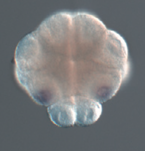
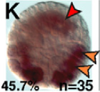
112-cell stage wild type embryos probed for Ci- Tbx6b expression.
In the second picture, the notochord and its precursors are outlined by a dashed red line.

Original Annotation : Ci-Tbx6b is expressed at the 112-cell stage in muscle precursors (orange arrowheads).

Stained molecule KH2012:KH.S654.3 (TBX2; TBX4; TBX6)
Stained region(s) B7.5 cell pair - B8.15 cell pair - B8.16 cell pair - b8.17 cell pair - B8.7 cell pair - B8.8 cell pair -

C. robusta formely Ciona int. type A, Stage 10 (112-cell)

Read more…

112-cell stage embryos probed for Ci-Tbx6b expression following the injection of pCi-Bra::Ci-Tbx6b construct.
The notochord and its precursors are outlined by a dashed red line.

Original Annotation : Embryos electroporated with the Bra>Tbx6b construct expressed Ci-Tbx6b in both the tail muscle (the endogenous gene) and the notochord (the transgene) at the 110-cell stage.

Stained molecule KH2012:KH.S654.3 (TBX2; TBX4; TBX6)
Stained region(s) A8.13 cell pair - A8.14 cell pair - A8.5 cell pair - A8.6 cell pair - B7.5 cell pair - B8.15 cell pair - B8.16 cell pair - B8.6 cell pair - B8.7 cell pair - B8.8 cell pair -

C. robusta formely Ciona int. type A, Stage 10 (112-cell)

Read more…

112-cell stage embryos probed for Ci- Tbx6b expression following the electroporation of pBra::macho1 construct.
The first picture is a low-magnification microphotograph of embryos.
The percentages of transgenic embryos displaying the patterns shown, and the total number of embryos scored, are indicated at the bottom of each panel.

Original Annotation : Ci-Tbx6b is expressed at the 112-cell stage in muscle precursors (I; orange arrowheads). Ectopic expression of Ci-Tbx6b is detected in the notochord of 112-cell-stage Bra>macho transgenics (red arrowhead).

Stained molecule KH2012:KH.S654.3 (TBX2; TBX4; TBX6)
Stained region(s) A8.13 cell pair - A8.14 cell pair - A8.5 cell pair - A8.6 cell pair - B7.5 cell pair - B8.15 cell pair - B8.16 cell pair - b8.17 cell pair - B8.6 cell pair - B8.7 cell pair - B8.8 cell pair -

C. robusta formely Ciona int. type A, Stage 10 (112-cell)

Read more…

112-cell stage embryos probed for Ci-Tbx6b expression following the electroporation of pBra::Tbx6c construct.
The percentages of transgenic embryos displaying the patterns shown, and the total number of embryos scored, are indicated at the bottom of each panel.

Original Annotation : Ci-Tbx6b is expressed at the 112-cell stage in muscle precursors (orange arrowheads). Ectopic notochord staining is indicated by red arrowheads.

Stained molecule KH2012:KH.S654.3 (TBX2; TBX4; TBX6)
Stained region(s) A8.13 cell pair - A8.14 cell pair - A8.5 cell pair - A8.6 cell pair - B7.5 cell pair - B8.15 cell pair - B8.16 cell pair - b8.17 cell pair - B8.6 cell pair - B8.7 cell pair - B8.8 cell pair -

C. robusta formely Ciona int. type A, Stage 10 (112-cell)

Read more…

Conversion to Aniseed format of data from Yutaka Satou's gene expression profiles of transcription factors and signalling molecule ISH screen (Development. 2004 131:4047-58).

Original annotation :
Gene name :Tbx6b/c/d.
Expression pattern : B8.7, B8.8, B7.5, B8.15, B8.16.
Comment : The expression in the B8.15 and B8.16 is dispersed within cytoplasm and looked weak.

Stained molecule KH2012:KH.S654.3 (TBX2; TBX4; TBX6)
Stained region(s) B7.5 cell pair - B8.15 cell pair - B8.16 cell pair - B8.7 cell pair - B8.8 cell pair -

C. robusta formely Ciona int. type A, Stage 10 (112-cell)

Read more…

Vegetal pole view of a 110-cell stage embryo probed with Ci-Tbx6b expression.

Stained molecule KH2012:KH.S654.3 (TBX2; TBX4; TBX6)
Stained region(s) B7.5 cell pair - B8.15 cell pair - B8.16 cell pair - B8.7 cell pair - B8.8 cell pair -

C. robusta formely Ciona int. type A, Stage 10 (112-cell)

Read more…

Vegetal pole view of a 110-cell stage embryo probed with Ci-Tbx6b expression. Injection of Ci-macho1 MO prevents the expression of Ci-Tbx6b in the B7.5 cell pair and in the B8.15 cell contrary to the control where Ci-Tbx6b seems to be expressed in B7.5, B8.7, B8.8 and B8.15 cell pairs.

Stained molecule KH2012:KH.S654.3 (TBX2; TBX4; TBX6)
Stained region(s) B8.7 cell pair - B8.8 cell pair -

C. robusta formely Ciona int. type A, Stage 10 (112-cell)

Read more…

Vegetal pole view of a 110-cell stage embryo probed with Ci-Tbx6b expression. Injection of Ci-macho1 MO and Ci-ZicL MO prevents the expression of Ci-Tbx6b contrary to the control where Ci-Tbx6b seems to be expressed in B7.5, B8.7, B8.8 and B8.15 cell pairs.

Stained molecule KH2012:KH.S654.3 (TBX2; TBX4; TBX6)
Stained region(s) No expression -

C. robusta formely Ciona int. type A, Stage 10 (112-cell)

Read more…

Expression of Ci-Tbx6b was detected by whole-mount in situ hybridization. Vegetal view of a 110-cell stage embryo.

Aniseed annotation:
According to the picture and to the others data stored in ANISEED, it seems that there is also expression in B7.5 and B8.16 cell pairs.

Stained molecule KH2012:KH.S654.3 (TBX2; TBX4; TBX6)
Stained region(s) B7.5 cell pair - B8.15 cell pair - B8.16 cell pair - B8.7 cell pair - B8.8 cell pair -

C. robusta formely Ciona int. type A, Stage 10 (112-cell)

Read more…

112-cell stage embryos electroporated with the 2.4 kb wild-type Ci-Tbx6b CRM, region upstream of Ci-Tbx6b from -2339bp to -13bp.
The first picture is a schematic representation of the electroporated region. Green boxes represent putative Ci-Macho1-binding sites; black arrows indicate their orientations. The orange rectangle depicts the proximal region of the 2.4 kb fragment.
The percentages of transgenic embryos displaying the patterns shown, and the total number of embryos scored, are indicated at the bottom of each panel.

Original Annotation : At the 112-cell stage, lacZ expression was detected in descendants of B6.4 blastomeres and in the B8.7 and B8.8 pairs, which derive from the B6.2 pair.

Stained molecule KH2012:KH.S654.3 (TBX2; TBX4; TBX6)
Stained region(s) B8.15 cell pair - B8.16 cell pair - B8.7 cell pair - B8.8 cell pair -

C. robusta formely Ciona int. type A, Stage 10 (112-cell)

Read more…

112-cell stage embryos electroporated with the 2.4 kb CRM with mutations in all Ci-Macho1-binding sites.
The first picture is a schematic representation of the electroporated region. Green boxes represent putative Ci-Macho1-binding sites; black arrows indicate their orientations. The orange rectangle depicts the proximal region of the 2.4 kb fragment.
The percentages of transgenic embryos displaying the patterns shown, and the total number of embryos scored, are indicated at the bottom of each panel.

Original Annotation : No expression of the reporter at the 112-cell stage.

Stained molecule KH2012:KH.S654.3 (TBX2; TBX4; TBX6)
Stained region(s) No expression -

C. robusta formely Ciona int. type A, Stage 10 (112-cell)

Read more…

112-cell stage embryos electroporated with the 862 bp Ci-Tbx6b CRM, which does not contain the Ci-Macho1-binding sites.
The first picture is a schematic representation of the electroporated region. The orange rectangle depicts the proximal region of the 2.4 kb fragment.
The percentages of transgenic embryos displaying the patterns shown, and the total number of embryos scored, are indicated at the bottom of each panel.

Original Annotation : No detectable signal at the 112-cell stage.

Stained molecule KH2012:KH.S654.3 (TBX2; TBX4; TBX6)
Stained region(s) No expression -

C. robusta formely Ciona int. type A, Stage 10 (112-cell)

Read more…

112-cell stage embryos electroporated with the fragment 266bp Ci-Tbx6b CRM containing the region upstream of Ci-Tbx6b from -145bp to -13bp upstream of basal promoter of Ci-Tbx6b.
The first picture is a schematic representation of the electroporated region.
The 266 bp proximal region of the Ci-Tbx6b CRM contains putative binding sites for CREB (blue diamond), T-box (red oval) and bHLH ("AC"-core E-box; pink triangle) transcription factors.
The percentages of transgenic embryos displaying the patterns shown, and the total number of embryos scored, are indicated at the bottom of each panel.

Original Annotation : The majority of the embryos electroporated with the 266 bp construct did not show staining at the 112-cell stage.

Stained molecule KH2012:KH.S654.3 (TBX2; TBX4; TBX6)
Stained region(s) No expression -

C. robusta formely Ciona int. type A, Stage 10 (112-cell)

Read more…

Embryos electroporated with either the distal muscle CRM and fixed at the 112-cell stage.
The distal region contains two E-boxes .

Original Annotation : In the majority of the embryos analyzed, high levels of lacZ transcripts were predominantly detected in only one pair of muscle precursors although a few embryos also showed a faint signal in additional muscle precursors (orange arrowheads), including the mixed-lineage A8.16 blastomeres (orange and blue arrowhead).

Stained molecule KH2012:KH.S654.3 (TBX2; TBX4; TBX6)
Stained region(s) A8.16* - B8.15 cell pair - B8.7 cell pair - B8.8 cell pair -

C. robusta formely Ciona int. type A, Stage 12 (mid gastrula)

Read more…

Vegetal view of a gastrula stage embryo probed for Ci-Tbx6b expression.

Stained molecule KH2012:KH.S654.3 (TBX2; TBX4; TBX6)
Stained region(s) tail muscles -

C. robusta formely Ciona int. type A, Stage 12 (mid gastrula)

Read more…

Vegetal view of a gastrula stage 5-aza-dC- treated embryo probed for Ci-Tbx6b expression.

Original annotation:
Appearance of the Ci-Tbx6b mRNAs was retarded.

Stained molecule KH2012:KH.S654.3 (TBX2; TBX4; TBX6)
Stained region(s) tail muscles -

C. robusta formely Ciona int. type A, Stage 13 (late gastrula)

Read more…

Late gastrula embryos probed for Ci-Tbx6b expression.
The notochord and its precursors are outlined by a dashed red line.

Original Annotation : Wild-type embryos show expression of Ci-Tbx6b in muscle precursors at the gastrula stage.

Stained molecule KH2012:KH.S654.3 (TBX2; TBX4; TBX6)
Stained region(s) tail muscles -

C. robusta formely Ciona int. type A, Stage 13 (late gastrula)

Read more…

Late gastrula embryos probed for Ci-Tbx6b expression following the injection of pCi-Bra::Ci-Tbx6b.
The notochord and its precursors are outlined by a dashed red line.

Original Annotation : Embryos electroporated with the Bra>Tbx6b construct expressed Ci-Tbx6b in both the tail muscle (the endogenous gene) and the notochord (the transgene) at the gastrula stage.

Stained molecule KH2012:KH.S654.3 (TBX2; TBX4; TBX6)
Stained region(s) notochord - tail muscles -

C. robusta formely Ciona int. type A, Stage 13 (late gastrula)

Read more…

Conversion to Aniseed format of data from Yutaka Satou's gene expression profiles of transcription factors and signalling molecule ISH screen (Development. 2004 131:4047-58).

Original annotation :
Gene name :Tbx6b/c/d.
Expression pattern : muscle

Stained molecule KH2012:KH.S654.3 (TBX2; TBX4; TBX6)
Stained region(s) tail muscles -

C. robusta formely Ciona int. type A, Stage 13 (late gastrula)

Read more…

Expression of Ci-Tbx6b was detected by whole-mount in situ hybridization. View of a late gastrula stage embryo.

Stained molecule KH2012:KH.S654.3 (TBX2; TBX4; TBX6)
Stained region(s) tail muscles -

C. robusta formely Ciona int. type A, Stage 13 (late gastrula)

Read more…

Vegetal pole view of a late tailbud stage wild type embryo probed for Ci-Tbx6b expression. This picture shows expression in the rowI/column4 (A9.31) an din the tail muscles. Numbers indicate any expression over the total number of embryos analysed. Nuclei are labeled with Hoechst (white) to allow the easy identification of individual cells.

Stained molecule KH2012:KH.S654.3 (TBX2; TBX4; TBX6)
Stained region(s) tail muscles -

C. robusta formely Ciona int. type A, Stage 13 (late gastrula)

Read more…

Vegetal pole view of a late tailbud stage embryo probed for Ci-Tbx6b expression in which Nodal expression has been blocked by SB431542. Inhibition of Nodal signaling inhibited ci-Tbx6b expression in A9.31 cell pair. Numbers indicate any expression over the total number of embryos analysed.

Stained molecule KH2012:KH.S654.3 (TBX2; TBX4; TBX6)
Stained region(s) primary muscle lineage -

C. robusta formely Ciona int. type A, Stage 13 (late gastrula)

Read more…

Vegetal pole view of a late tailbud stage embryo probed for Ci-Tbx6b expression in which Nodal expression has been blocked by Nodal-Mo. Inhibition of Nodal signaling inhibited ci-Tbx6b expression in A9.31 cell pair. Numbers indicate any expression over the total number of embryos analysed.

Stained molecule KH2012:KH.S654.3 (TBX2; TBX4; TBX6)
Stained region(s) primary muscle lineage -

C. robusta formely Ciona int. type A, Stage 13 (late gastrula)

Read more…

Vegetal view of a late gastrula wild type embryo probed for Ci-tbx6b expression.

Original Annotation : At this stage the Ci-tbx6b staining is detected in the rowI column 4 (A9.31) and in the tail muscle.

Stained molecule KH2012:KH.S654.3 (TBX2; TBX4; TBX6)
Stained region(s) tail muscles -

C. robusta formely Ciona int. type A, Stage 13 (late gastrula)

Read more…

Vegetal view of a late gastrula stage embryo probed for Ci-tbx6b expression following DAPT injection.
Graph shows the percentage of embryos with ectopic, strong or weak Ci-tbx6b expression. ('Weak' means at least one cell with downregulated expression, 'strong' means at least one A-line precursor cell with strong expression).

Original Annotation : There is a loss of the expression of Ci-tbx6b in A9.31 in neural plate after the inhibition of Delta2/Notch signaling (due to DAPT).

Stained molecule KH2012:KH.S654.3 (TBX2; TBX4; TBX6)
Stained region(s) tail muscles -

C. robusta formely Ciona int. type A, Stage 13 (late gastrula)

Read more…

Vegetal view of a late gastrula stage embryo probed for Ci-tbx6b expression following the injection of a Delta2-MO.
Graph shows the percentage of embryos with ectopic, strong or weak Ci-tbx6b expression. ('Weak' means at least one cell with downregulated expression, 'strong' means at least one A-line precursor cell with strong expression).

Original Annotation : There is a loss of the expression of Ci-tbx6b in A9.31 in neural plate after the inhibition of Delta2/Notch signaling (due to Del2-MO).

Stained molecule KH2012:KH.S654.3 (TBX2; TBX4; TBX6)
Stained region(s) tail muscles -

C. robusta formely Ciona int. type A, Stage 13 (late gastrula)

Read more…

Graph showing the percentage of embryos with strong or weak Ci-tbx6b expression following the injection of dominant negative of Delta2, DnDel2. 'Weak' means at least one cell with downregulated expression, 'strong' means at least one A-line precursor cell with strong expression.

Original Annotation : Ci-tbx6b expression was weak following the inhibition of Delta2/Notch signaling.

Stained molecule KH2012:KH.S654.3 (TBX2; TBX4; TBX6)
Stained region(s) tail muscles -

C. robusta formely Ciona int. type A, Stage 14 (early neurula)

Read more…

Early neurula embryos electroporated with the 2.4 kb CRM with mutations in all Ci-Macho1-binding sites.
The first picture is a schematic representation of the electroporated region. Green boxes represent putative Ci-Macho1-binding sites; black arrows indicate their orientations. The orange rectangle depicts the proximal region of the 2.4 kb fragment.
The percentages of transgenic embryos displaying the patterns shown, and the total number of embryos scored, are indicated at the bottom of each panel.

Original Annotation : The vast majority of embryos carrying the 2.4 kb CRM with mutations in all Ci-Macho1-binding sites displayed active transcription in the posterior-most muscle cells only at the neurula stage.

Stained molecule KH2012:KH.S654.3 (TBX2; TBX4; TBX6)
Stained region(s) primary muscle lineage -

C. robusta formely Ciona int. type A, Stage 14 (early neurula)

Read more…

Early neurula embryos electroporated with the 862 bp Ci-Tbx6b CRM, which does not contain the Ci-Macho1-binding sites.
The first picture is a schematic representation of the electroporated region. The orange rectangle depicts the proximal region of the 2.4 kb fragment.
The percentages of transgenic embryos displaying the patterns shown, and the total number of embryos scored, are indicated at the bottom of each panel.

Original Annotation : LacZ expression became detectable by the early neurula stage in only the posterior-most muscle cells (note that the asymmetrical expression is due to mosaic incorporation of the transgene).

Stained molecule KH2012:KH.S654.3 (TBX2; TBX4; TBX6)
Stained region(s) primary muscle lineage -

C. robusta formely Ciona int. type A, Stage 14 (early neurula)

Read more…

Early neurula stage embryos electroporated with the fragment 266bp Ci-Tbx6b CRM containing the region upstream of Ci-Tbx6b from -145bp to -13bp upstream of basal promoter of Ci-Tbx6b.
The first picture is a schematic representation of the electroporated region.
The 266 bp proximal region of the Ci-Tbx6b CRM contains putative binding sites for CREB (blue diamond), T-box (red oval) and bHLH ("AC"-core E-box; pink triangle) transcription factors.
The percentages of transgenic embryos displaying the patterns shown, and the total number of embryos scored, are indicated at the bottom of each panel.

Original Annotation : By the early neurula stage, 87% of the embryos showed strong staining in muscle cells.

Stained molecule KH2012:KH.S654.3 (TBX2; TBX4; TBX6)
Stained region(s) tail muscles -

C. robusta formely Ciona int. type A, Stage 14 (early neurula)

Read more…

Embryos electroporated with either the distal muscle CRM and fixed at the early neurula stage.
The distal region contains two E-boxes .

Original Annotation : Consistent with this early pattern, at the early neurula stage beta-galactosidase accumulation was detected only in a small subset of muscle and mesenchyme precursors.

Stained molecule KH2012:KH.S654.3 (TBX2; TBX4; TBX6)
Stained region(s) tail muscles -

C. robusta formely Ciona int. type A, Stage 14 (early neurula)

Read more…

Early neurula embryos electroporated with the 2.4 kb wild-type Ci-Tbx6b CRM, region upstream of Ci-Tbx6b from -2339bp to -13bp.
The first picture is a schematic representation of the electroporated region. Green boxes represent putative Ci-Macho1-binding sites; black arrows indicate their orientations. The orange rectangle depicts the proximal region of the 2.4 kb fragment.
The percentages of transgenic embryos displaying the patterns shown, and the total number of embryos scored, are indicated at the bottom of each panel.

Original Annotation : In early neurulae, lacZ expression was detected only in the posterior-most muscle cells. This pattern faithfully recapitulates the expression of Ci-Tbx6b.

Stained molecule KH2012:KH.S654.3 (TBX2; TBX4; TBX6)
Stained region(s) primary muscle lineage -

C. robusta formely Ciona int. type A, Stage 15 (mid neurula)

Read more…

Dorsal view of a neurula stage embryo probed for Ci-Tbx6b expression.

Stained molecule KH2012:KH.S654.3 (TBX2; TBX4; TBX6)
Stained region(s) tail muscles -

C. robusta formely Ciona int. type A, Stage 15 (mid neurula)

Read more…

Dorsal view of a neurula 5-aza-dC- treated embryo probed for Ci-Tbx6b expression.

Original annotation:
Appearance of the Ci-Tbx6b mRNAs was retarded.

Stained molecule KH2012:KH.S654.3 (TBX2; TBX4; TBX6)
Stained region(s) No expression -

C. robusta formely Ciona int. type A, Stage 15 (mid neurula)

Read more…

Conversion to Aniseed format of data from Yutaka Satous gene expression profiles of transcription factors and signalling molecule ISH screen (Development. 2004 131:4047-58).

Original annotation :
Gene name :Tbx6b/c/d.
Expression pattern : muscle.
Comment : The primary muscle was stained lightly and the secondary muscle was stained deeply.

Stained molecule KH2012:KH.S654.3 (TBX2; TBX4; TBX6)
Stained region(s) tail muscles -

C. robusta formely Ciona int. type A, Stage 15 (mid neurula)

Read more…

Expression of Ci-Tbx6b was detected by whole-mount in situ hybridization. View of a mid-neurula stage embryo.

Stained molecule KH2012:KH.S654.3 (TBX2; TBX4; TBX6)
Stained region(s) tail muscles -

C. robusta formely Ciona int. type A, Stage 19 (early tailbud I)

Read more…

Conversion to Aniseed format of data from Yutaka Satou's gene expression profiles of transcription factors and signalling molecule ISH screen (Development. 2004 131:4047-58).

Original annotation :
Gene name :Tbx6b/c/d.
Expression pattern : No distinct zygotic signal was detected

Stained molecule KH2012:KH.S654.3 (TBX2; TBX4; TBX6)
Stained region(s) No expression -

C. robusta formely Ciona int. type A, Stage 19 (early tailbud I)

Read more…

Embryos electroporated with either the distal muscle CRM and fixed at the early tailbud stage.
The distal region contains two E-boxes .

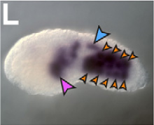
Original Annotation : By the early tailbud stage, the activity of the CRM had considerably expanded, to encompass virtually all muscle cells (orange arrowheads), as well as part of the nerve cord (blue arrowheads) and mesenchyme cells (pink arrowheads).

Stained molecule KH2012:KH.S654.3 (TBX2; TBX4; TBX6)
Stained region(s) tail muscles - tail nerve cord -

C. robusta formely Ciona int. type A, Stage 21 (mid tailbud I)

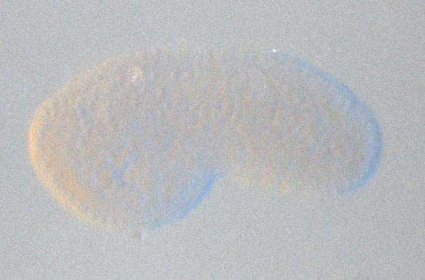
Read more…

Mid tailbud embryos probed for Ci-Tbx6b expression. Embryos are oriented with anterior to the left, dorsal up.
In the second picture, the notochord and its precursors are outlined by a dashed red line.

Original Annotation : Ci-Tbx6b is expressed at the 112-cell stage in muscle precursors but is no longer expressed by the early tailbud stage.

Stained molecule KH2012:KH.S654.3 (TBX2; TBX4; TBX6)
Stained region(s) No expression -

C. robusta formely Ciona int. type A, Stage 21 (mid tailbud I)

Read more…

Mid tailbud embryos probed for Ci-Tbx6b expression following the injection of pCi-Bra::Ci-Tbx6b construct.
The notochord and its precursors are outlined by a dashed red line.

Original Annotation : Embryos electroporated with the Bra>Tbx6b construct expressed Ci-Tbx6b at the tailbud stage, only in the notochord, expression directed by the transgene was detected.

Stained molecule KH2012:KH.S654.3 (TBX2; TBX4; TBX6)
Stained region(s) notochord -

C. robusta formely Ciona int. type A, Stage 21 (mid tailbud I)

Read more…

Mid tailbud stage embryos probed for Ci- Tbx6b expression following the electroporation of pBra::macho1 construct.
The first picture is a low-magnification microphotograph of embryos.
The percentages of transgenic embryos displaying the patterns shown, and the total number of embryos scored, are indicated at the bottom of each panel.

Original Annotation : Mid-tailbud Bra>macho embryos show ectopic expression of Ci-Tbx6b only in mesenchyme (pink arrowhead).

Stained molecule KH2012:KH.S654.3 (TBX2; TBX4; TBX6)
Stained region(s)

C. robusta formely Ciona int. type A, Stage 21 (mid tailbud I)

Read more…

Conversion to Aniseed format of data from Yutaka Satou's gene expression profiles of transcription factors and signalling molecule ISH screen (Development. 2004 131:4047-58).

Original annotation :
Gene name :Tbx6b/c/d.
Expression pattern : No distinct zygotic signal was detected

Stained molecule KH2012:KH.S654.3 (TBX2; TBX4; TBX6)
Stained region(s) No expression -

C. robusta formely Ciona int. type A, Stage 21 (mid tailbud I)

Read more…

A 112 bp subfragment of the 266 bp CRM, containing the three putative binding sites(CREB (blue diamond), T-box (red oval) and E-box (pink triangle)) was cloned upstream of the Ci-FoxA-a basal promoter (pink box).
Inset show transgenic embryos fixed at the neurula stage.
The number of embryos scored for each construct is shown in white.
Low-magnification image of mid-tailbud embryos electroporated with the wild-type 112 bp Ci-Tbx6b CRM.

Aniseed Annotation : The reporter was expressed in the muscle and mesenchyme.

Stained molecule KH2012:KH.S654.3 (TBX2; TBX4; TBX6)
Stained region(s) tail muscles -

C. robusta formely Ciona int. type A, Stage 21 (mid tailbud I)

Read more…

A 112 bp subfragment of the 266 bp CRM, containing the three putative binding sites(CREB (blue diamond), T-box (red oval) and E-box (pink triangle)) was cloned upstream of the Ci-FoxA-a basal promoter (pink box). Here the T-box site was mutated (white crossed oval).
Inset show transgenic embryos fixed at the neurula stage.
The number of embryos scored for each construct is shown in white.

Low-magnification image of mid-tailbud embryos electroporated with the wild-type 112 bp Ci-Tbx6b CRM.

Original Annotation : When the T-box binding site was mutated, a considerable reduction was observed not only in the intensity of the muscle staining but also in the percentage of embryos showing activation of the reporter gene.

Stained molecule KH2012:KH.S654.3 (TBX2; TBX4; TBX6)
Stained region(s) No expression -

C. robusta formely Ciona int. type A, Stage 21 (mid tailbud I)

Read more…

Microphotographs of X-Gal-stained Ciona embryos electroporated with the 400 bp region, cloned upstream of the FoxA-a basal promoter (pink box in the schematics). The distal region contains two E-boxes (pink triangles).

Reporter expression was detected in muscle (orange arrowheads) and in the CNS (blue arrowheads).

Stained molecule KH2012:KH.S654.3 (TBX2; TBX4; TBX6)
Stained region(s) central nervous system - tail muscles -

C. robusta formely Ciona int. type A, Stage 21 (mid tailbud I)

Read more…

Embryos electroporated with either the distal muscle CRM and fixed at the mid tailbud stage.
The distal region contains two E-boxes .

Original Annotation : The CRM remained active in most muscle cells (orange arrowheads) through the mid-tailbud stage.

Stained molecule KH2012:KH.S654.3 (TBX2; TBX4; TBX6)
Stained region(s) tail muscles -

C. robusta formely Ciona int. type A, Stage 21 (mid tailbud I)

Read more…

Early neurula embryos electroporated with the 2.4 kb wild-type Ci-Tbx6b CRM, region upstream of Ci-Tbx6b from -2339bp to -13bp.
The first picture is a schematic representation of the electroporated region. Green boxes represent putative Ci-Macho1-binding sites; black arrows indicate their orientations. The orange rectangle depicts the proximal region of the 2.4 kb fragment.
The second picture is a low-magnification microphotographs of Ciona intestinalis embryos electroporated with either the 2.4 kb Ci-Tbx6b CRM.

Original Annotation : Orange and purple arrowheads indicate muscle and mesenchyme cells, respectively.

Stained molecule KH2012:KH.S654.3 (TBX2; TBX4; TBX6)
Stained region(s) tail muscles -

C. robusta formely Ciona int. type A, Stage 21 (mid tailbud I)

Read more…

Embryos electroporated with a fragment of 456bp (named 504bp in the paper) upstream of Ci-Tbx6b, cloned upstream of Ci-FoxAa, and probed for reporter expression in muscle.
Red dashed lines represent the minimal 112bp sequence driving muscle expression.

Expression was detected in muscle.

Stained molecule KH2012:KH.S654.3 (TBX2; TBX4; TBX6)
Stained region(s) tail muscles -